Copyright
©The Author(s) 2021.
World J Clin Cases. May 16, 2021; 9(14): 3449-3457
Published online May 16, 2021. doi: 10.12998/wjcc.v9.i14.3449
Published online May 16, 2021. doi: 10.12998/wjcc.v9.i14.3449
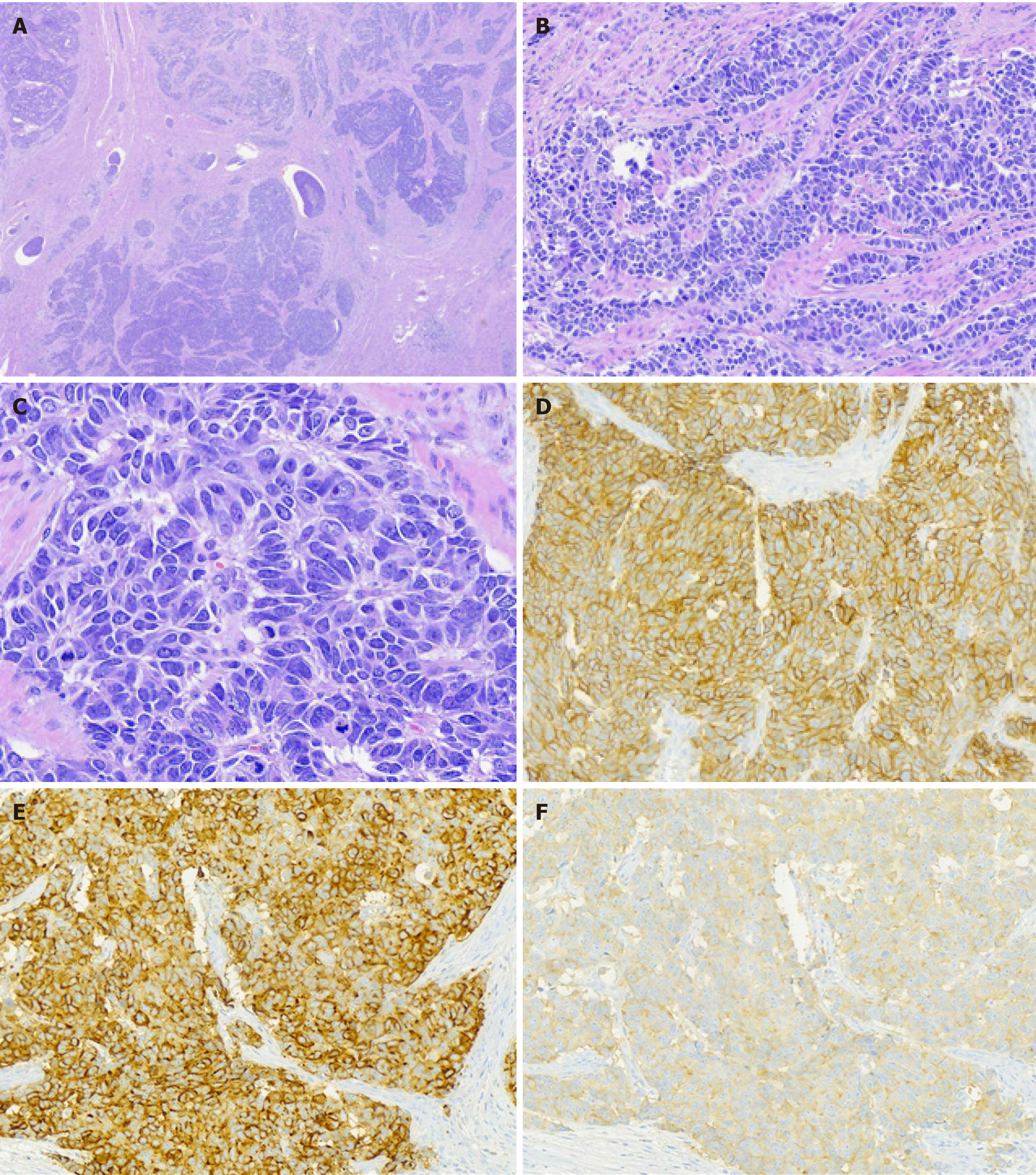
Figure 2 Histology and immunohistochemistry.
A: Microscopically, tumors showed an organoid pattern of growth with tumor embolism (hematoxylin and eosin, × 40); B: Trabeculae-like pattern (hematoxylin and eosin, × 200); C: Rosette-like pattern with “salt and pepper” chromatin (hematoxylin and eosin, × 400); D-F: Immunohistochemistry revealed positivity for CD56 (D, membranous), synaptophysin (E, cytoplasmic), and chromogranin A (F, cytoplasmic).
- Citation: Du R, Jiang F, Wang ZY, Kang YQ, Wang XY, Du Y. Pure large cell neuroendocrine carcinoma originating from the endometrium: A case report. World J Clin Cases 2021; 9(14): 3449-3457
- URL: https://www.wjgnet.com/2307-8960/full/v9/i14/3449.htm
- DOI: https://dx.doi.org/10.12998/wjcc.v9.i14.3449


