Copyright
©The Author(s) 2021.
World J Clin Cases. May 16, 2021; 9(14): 3411-3417
Published online May 16, 2021. doi: 10.12998/wjcc.v9.i14.3411
Published online May 16, 2021. doi: 10.12998/wjcc.v9.i14.3411
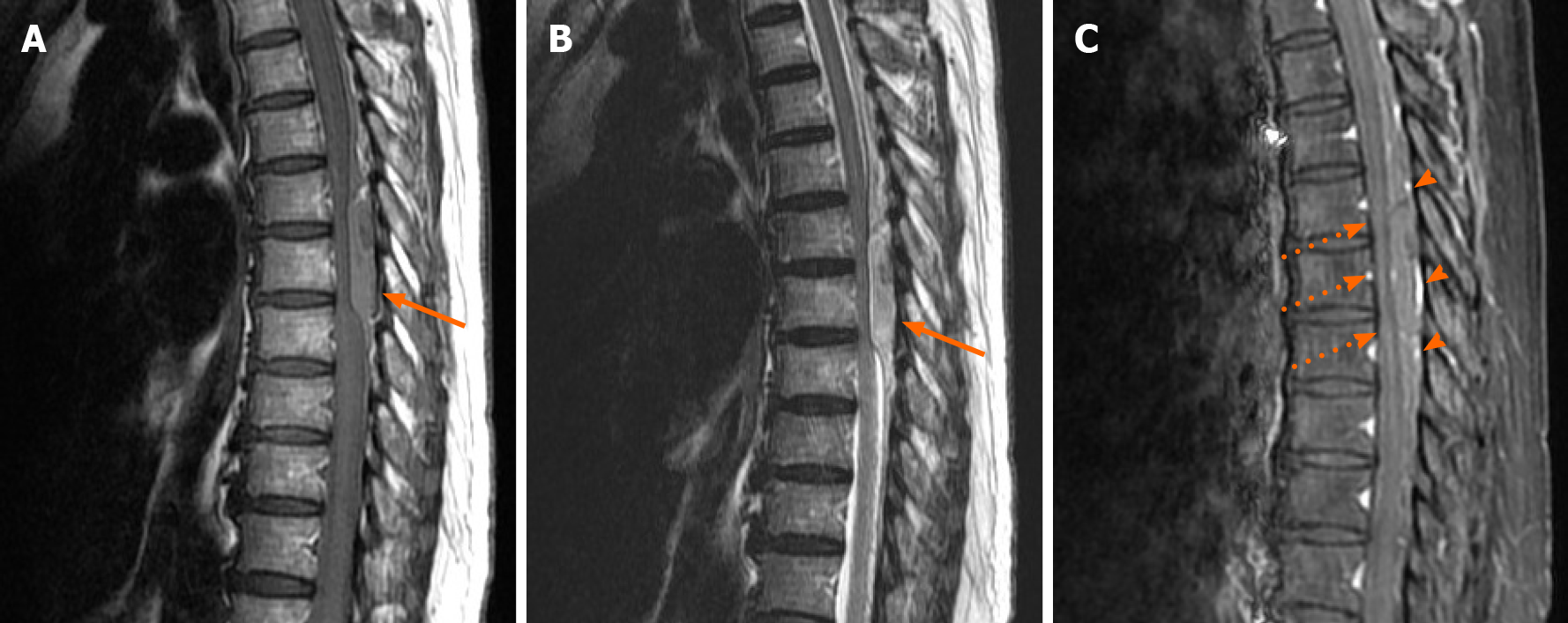
Figure 2 Preoperative spinal magnetic resonance imaging demonstrating an intraspinal epidural hematoma at the T4 to the T8 levels (arrows).
A: Sagittal T1-weighted image (T1WI); B: Sagittal T2-weighted image; C: Sagittal T1WI with enhancement showing mildly thin peripheral enhancement (arrowheads) and the spinal cord is compressed and flattened (dashed thin arrows).
- Citation: Chia KJ, Lin LH, Sung MT, Su TM, Huang JF, Lee HL, Sung WW, Lee TH. Acute spontaneous thoracic epidural hematoma associated with intraspinal lymphangioma: A case report. World J Clin Cases 2021; 9(14): 3411-3417
- URL: https://www.wjgnet.com/2307-8960/full/v9/i14/3411.htm
- DOI: https://dx.doi.org/10.12998/wjcc.v9.i14.3411


