Copyright
©The Author(s) 2021.
World J Clin Cases. May 16, 2021; 9(14): 3403-3410
Published online May 16, 2021. doi: 10.12998/wjcc.v9.i14.3403
Published online May 16, 2021. doi: 10.12998/wjcc.v9.i14.3403
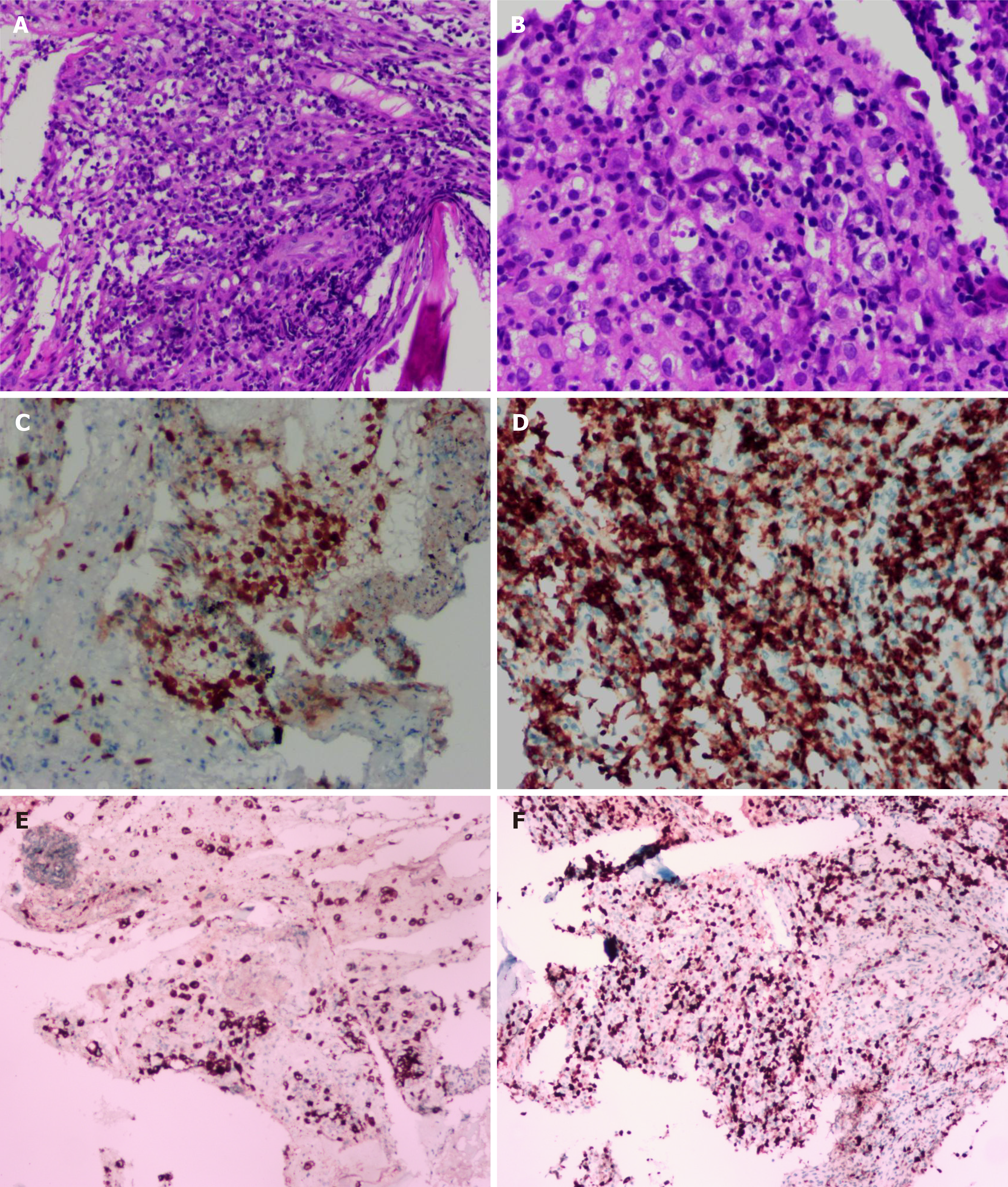
Figure 3 Histopathological microphotograph of primary bone anaplastic large-cell lymphoma.
A: The lesions of the vertebral body were infiltrated by pleomorphic tumor cells which have scanty cytoplasm and hyperchromatic nuclei (magnification × 100); B: The neoplastic cells exhibited small- to medium-sized, irregular nuclei and abundant clear cytoplasm. Hallmark cells (horseshoe-shaped or doughnut cells) were present and Reed-Sternberg cell-like cell were also noted (magnification × 400); C-F: On immunohistochemistry, the large atypical cells showed positive expression of anaplastic lymphoma kinase (C), CD3 (D), and CD30 (E), and the Ki67 index was almost 60% (F).
- Citation: Zheng W, Yin QQ, Hui TC, Wu WH, Wu QQ, Huang HJ, Chen MJ, Yan R, Huang YC, Pan HY. Primary bone anaplastic lymphoma kinase positive anaplastic large-cell lymphoma: A case report and review of the literature . World J Clin Cases 2021; 9(14): 3403-3410
- URL: https://www.wjgnet.com/2307-8960/full/v9/i14/3403.htm
- DOI: https://dx.doi.org/10.12998/wjcc.v9.i14.3403


