Copyright
©The Author(s) 2021.
World J Clin Cases. May 16, 2021; 9(14): 3365-3371
Published online May 16, 2021. doi: 10.12998/wjcc.v9.i14.3365
Published online May 16, 2021. doi: 10.12998/wjcc.v9.i14.3365
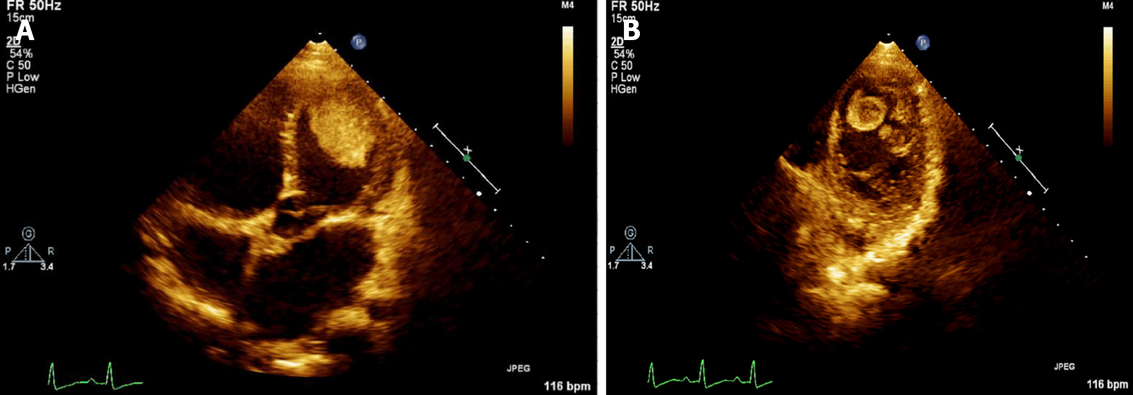
Figure 1 False color imaging of thrombi using two-dimensional echocardiography.
A: Apical four-chamber view shows a large thrombus near the apex of the lateral wall of the left ventricle; B: Two thrombi were also detected at the apex of the left ventricle.
- Citation: Sun LJ, Li Y, Qiao W, Yu JH, Ren WD. Incremental value of three-dimensional and contrast echocardiography in the evaluation of endocardial fibroelastosis and multiple cardiovascular thrombi: A case report. World J Clin Cases 2021; 9(14): 3365-3371
- URL: https://www.wjgnet.com/2307-8960/full/v9/i14/3365.htm
- DOI: https://dx.doi.org/10.12998/wjcc.v9.i14.3365


