Copyright
©The Author(s) 2021.
World J Clin Cases. May 6, 2021; 9(13): 3185-3193
Published online May 6, 2021. doi: 10.12998/wjcc.v9.i13.3185
Published online May 6, 2021. doi: 10.12998/wjcc.v9.i13.3185
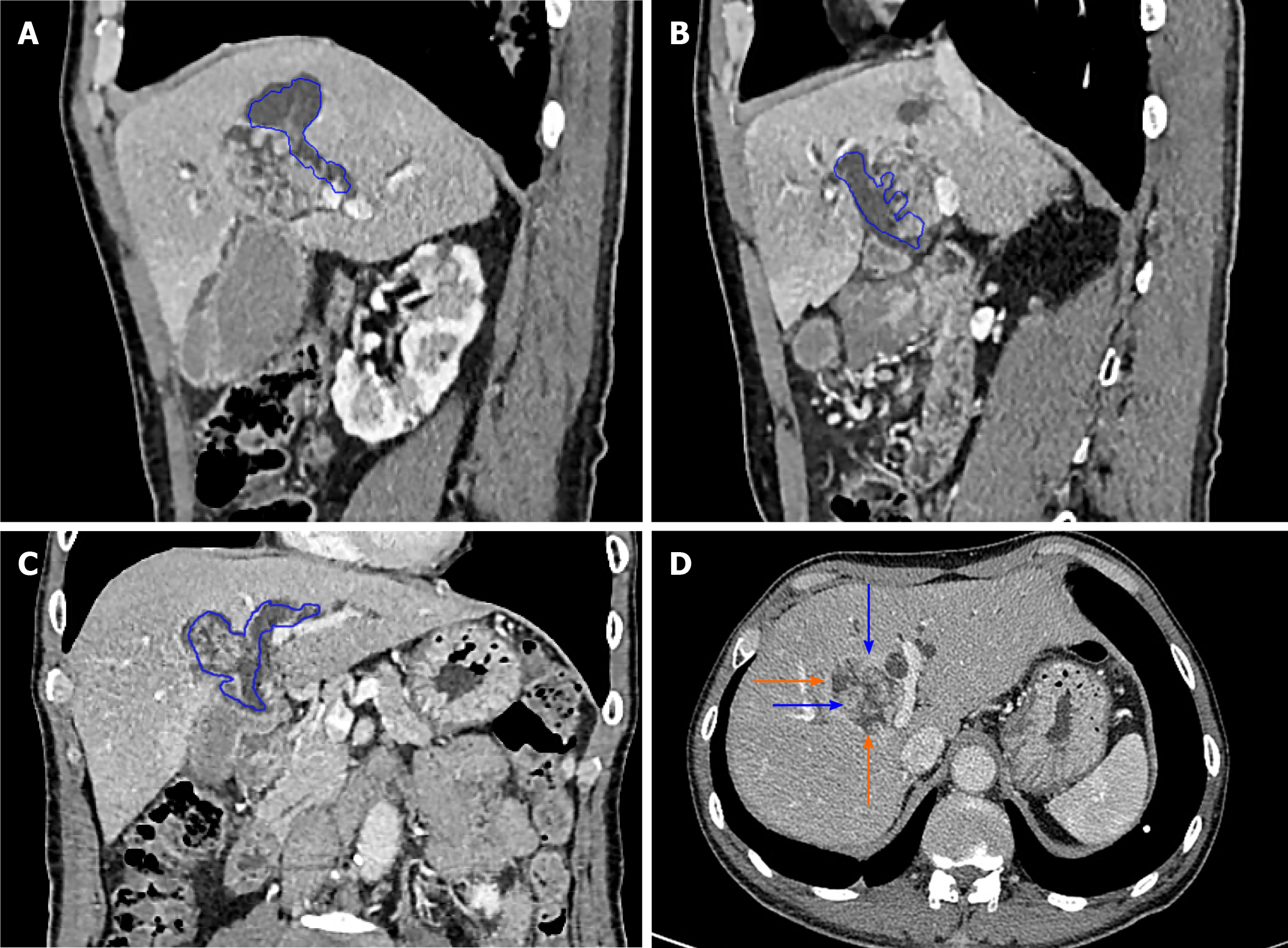
Figure 4 Intrahepatic bile duct papilloma whole abdominal plain scan and enhanced computed tomography scan.
A-C: Blue depicts that the tumour communicates with the bile duct, and bile duct dilatation can be seen; D: The orange arrow indicates a mucinous tumour, and the blue arrow indicates a mural nodule.
- Citation: Yi D, Zhao LJ, Ding XB, Wang TW, Liu SY. Clinical characteristics of intrahepatic biliary papilloma: A case report. World J Clin Cases 2021; 9(13): 3185-3193
- URL: https://www.wjgnet.com/2307-8960/full/v9/i13/3185.htm
- DOI: https://dx.doi.org/10.12998/wjcc.v9.i13.3185


