
















©The Author(s) 2021.
World J Clin Cases. May 6, 2021; 9(13): 3147-3156
Published online May 6, 2021. doi: 10.12998/wjcc.v9.i13.3147
Published online May 6, 2021. doi: 10.12998/wjcc.v9.i13.3147
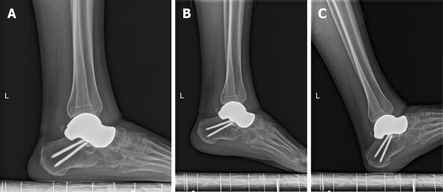
Figure 6 Postoperative radiographs (12 mo).
A: Lateral X-ray film of the ankle joint at 12 mo after operation; B: Extreme plantar flexion position; C: Extreme dorsiflexion position. The talus prosthesis was in place without displacement or subsidence, the surrounding bone was in good condition, and there was no instability or fracture around the prosthesis.
- Citation: Yang QD, Mu MD, Tao X, Tang KL. Three-dimensional printed talar prosthesis with biological function for giant cell tumor of the talus: A case report and review of the literature. World J Clin Cases 2021; 9(13): 3147-3156
- URL: https://www.wjgnet.com/2307-8960/full/v9/i13/3147.htm
- DOI: https://dx.doi.org/10.12998/wjcc.v9.i13.3147