Copyright
©The Author(s) 2021.
World J Clin Cases. May 6, 2021; 9(13): 3102-3113
Published online May 6, 2021. doi: 10.12998/wjcc.v9.i13.3102
Published online May 6, 2021. doi: 10.12998/wjcc.v9.i13.3102
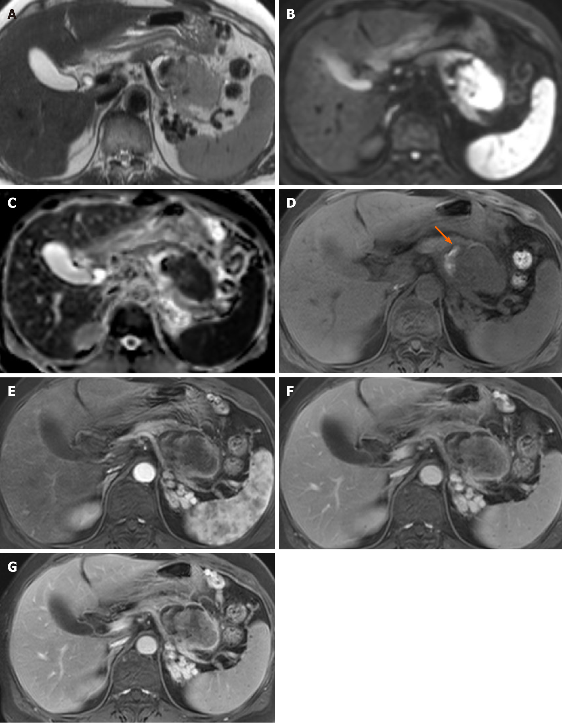
Figure 3 Magnetic resonance imaging findings.
A-D: An axial T2-weighted image showed an intermediate-to-high-signal intensity (SI) solid mass with peripheral cystic lesions in the pancreatic body (A); the solid portion in the mass showed diffusion restriction on the diffusion-weighted image (B); an apparent diffusion coefficient map (C); a small amount of intra-tumoral hemorrhage (arrow), showing high SI on an axial T1-weighted image (D) and low SI on a T2-weighted image was noted in the peripheral cystic portion (A); E-G: Axial contrast-enhanced dynamic T1-weighted images demonstrated significant peripheral progressive enhancement of the central solid portion.
- Citation: Lim HJ, Kang HS, Lee JE, Min JH, Shin KS, You SK, Kim KH. Sarcomatoid carcinoma of the pancreas — multimodality imaging findings with serial imaging follow-up: A case report and review of literature. World J Clin Cases 2021; 9(13): 3102-3113
- URL: https://www.wjgnet.com/2307-8960/full/v9/i13/3102.htm
- DOI: https://dx.doi.org/10.12998/wjcc.v9.i13.3102


