
















©The Author(s) 2021.
World J Clin Cases. Apr 26, 2021; 9(12): 2944-2950
Published online Apr 26, 2021. doi: 10.12998/wjcc.v9.i12.2944
Published online Apr 26, 2021. doi: 10.12998/wjcc.v9.i12.2944
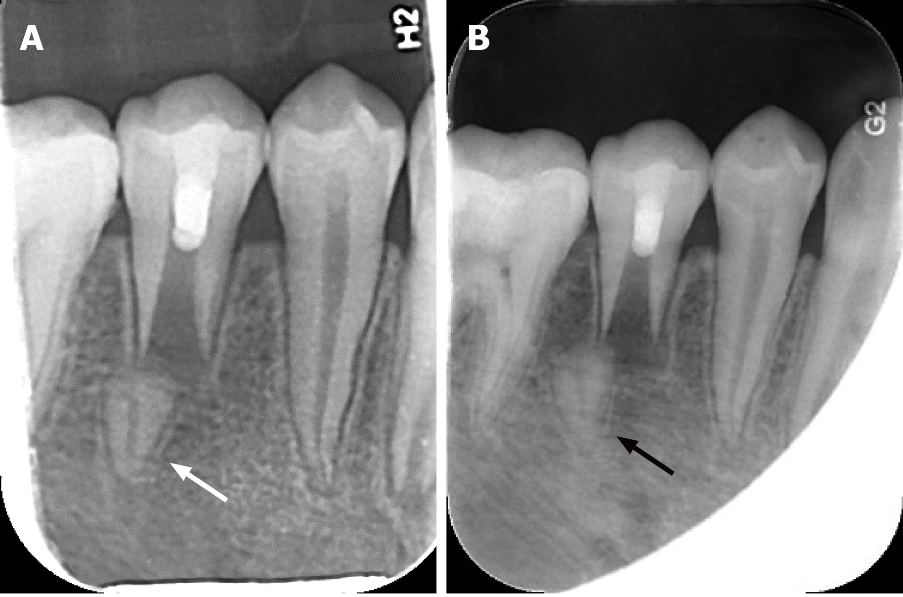
Figure 3 Periapical radiograph during the follow-up period.
A: Periapical radiograph at the 3 mo follow-up demonstrated complete resolution of the radiolucency and a drifting root tip (white arrow); B: Periapical radiograph at the 1 yr follow-up showed that the separated root tip (black arrow) was more distally drifted than before. The root length and dentin thickness of the main root did not increase.
- Citation: Wu ZF, Lu LJ, Zheng HY, Tu Y, Shi Y, Zhou ZH, Fang LX, Fu BP. Separated root tip formation associated with a fractured tubercle of dens evaginatus: A case report. World J Clin Cases 2021; 9(12): 2944-2950
- URL: https://www.wjgnet.com/2307-8960/full/v9/i12/2944.htm
- DOI: https://dx.doi.org/10.12998/wjcc.v9.i12.2944