Copyright
©The Author(s) 2021.
World J Clin Cases. Apr 26, 2021; 9(12): 2868-2873
Published online Apr 26, 2021. doi: 10.12998/wjcc.v9.i12.2868
Published online Apr 26, 2021. doi: 10.12998/wjcc.v9.i12.2868
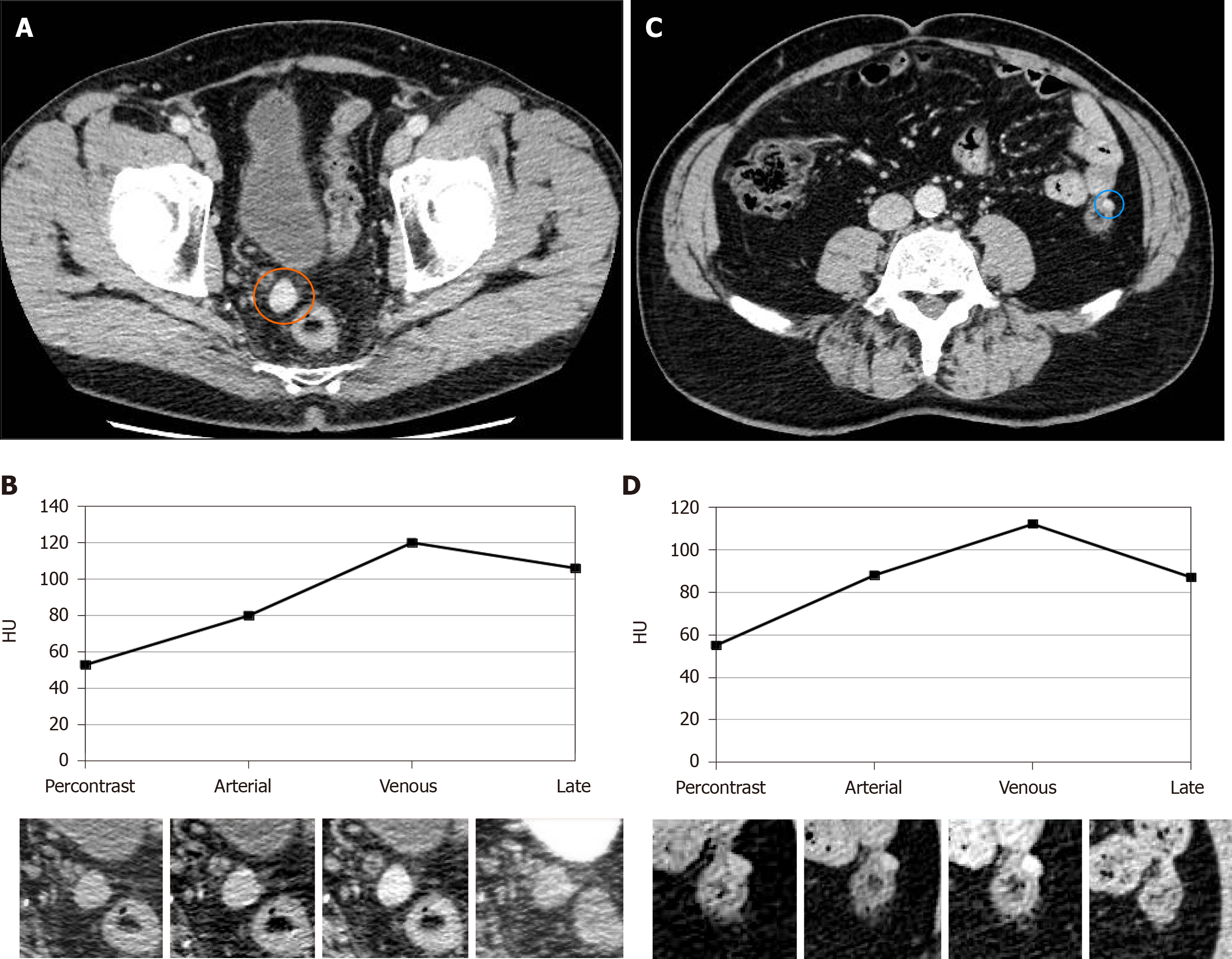
Figure 1 Computed tomography examination.
A: Multiphase contrast-enhanced computed tomography (CT) examination shows a rounded, smooth marginated nodule in the right recto-vesical space; B: Spleen-like vascularization, characterized by progressive contrast uptake in the arterial and venous phases and partial washout in the late phase; C and D: A further, smaller nodule with similar CT features can be seen adjacent to the left colon. HU: Hounsfield units.
- Citation: Tognarelli A, Faggioni L, Erba AP, Faviana P, Durante J, Manassero F, Selli C. Robotically assisted removal of pelvic splenosis fifty-six years after splenectomy: A case report. World J Clin Cases 2021; 9(12): 2868-2873
- URL: https://www.wjgnet.com/2307-8960/full/v9/i12/2868.htm
- DOI: https://dx.doi.org/10.12998/wjcc.v9.i12.2868


