Copyright
©The Author(s) 2021.
World J Clin Cases. Apr 26, 2021; 9(12): 2830-2837
Published online Apr 26, 2021. doi: 10.12998/wjcc.v9.i12.2830
Published online Apr 26, 2021. doi: 10.12998/wjcc.v9.i12.2830
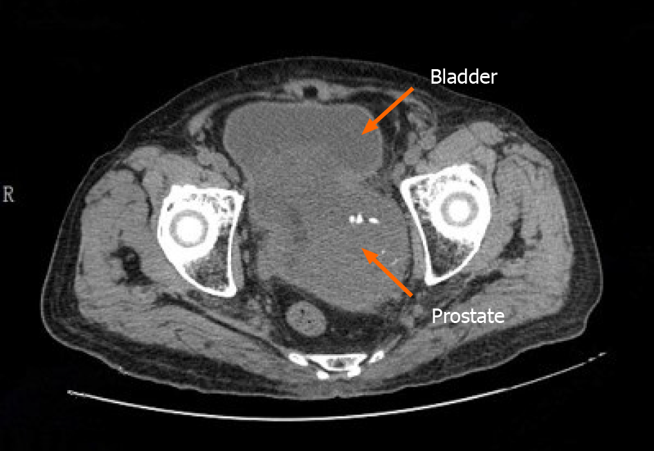
Figure 4 A follow-up pelvic computed tomography revealed that the prostate was enlarged in size with irregular morphology.
The prostate gland protruded locally to the bladder with uneven density. Patchy, low-density shadows and punctate calcification can also be seen in the prostate gland with an unclear boundary between the prostate and bilateral seminal vesicles gland.
- Citation: Zhao LW, Sun J, Wang YY, Hua RM, Tai SC, Wang K, Fan Y. Prostate stromal tumor with prostatic cysts after transurethral resection of the prostate: A case report. World J Clin Cases 2021; 9(12): 2830-2837
- URL: https://www.wjgnet.com/2307-8960/full/v9/i12/2830.htm
- DOI: https://dx.doi.org/10.12998/wjcc.v9.i12.2830


