Copyright
©The Author(s) 2021.
World J Clin Cases. Apr 26, 2021; 9(12): 2823-2829
Published online Apr 26, 2021. doi: 10.12998/wjcc.v9.i12.2823
Published online Apr 26, 2021. doi: 10.12998/wjcc.v9.i12.2823
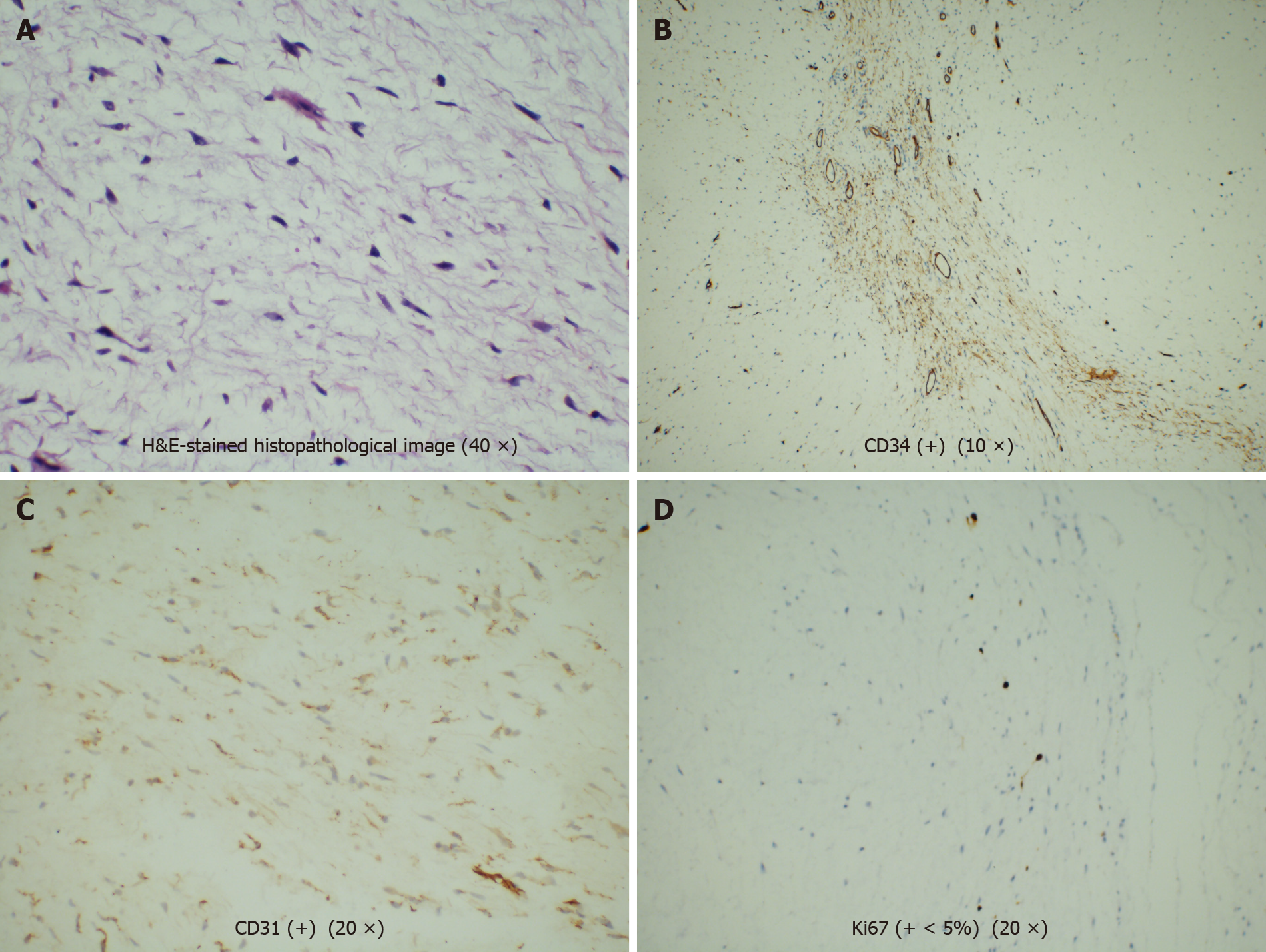
Figure 3 Hematoxylin & eosin-stained histopathological image (40 ×).
A: Hematoxylin & eosin (H&E)-stained histopathological image; B: Immunohistochemical staining of cluster of differentiation 34 (CD34) (+) (10 ×); C: Immunohistochemical staining of CD31 (+) (20 ×); D: Immunohistochemical staining of Ki67 (+ < 5%) (20 ×). The tumor had no envelope and showed expansive growth. It was composed of well-differentiated phyllodes mucinous tumors separated by fibroid tissues. There was no obvious atypia, mitosis, or necrosis. Part of the epithelium was missing, and mucosal ulcers were formed. The immunohistochemical results were as follows: Ki-67 (+ < 5%), CD31 (+), and CD34 (weak +).
- Citation: Yu TT, Yu H, Cui Y, Liu W, Cui XY, Wang X. Laryngeal myxoma: A case report. World J Clin Cases 2021; 9(12): 2823-2829
- URL: https://www.wjgnet.com/2307-8960/full/v9/i12/2823.htm
- DOI: https://dx.doi.org/10.12998/wjcc.v9.i12.2823


