Copyright
©The Author(s) 2021.
World J Clin Cases. Apr 26, 2021; 9(12): 2811-2815
Published online Apr 26, 2021. doi: 10.12998/wjcc.v9.i12.2811
Published online Apr 26, 2021. doi: 10.12998/wjcc.v9.i12.2811
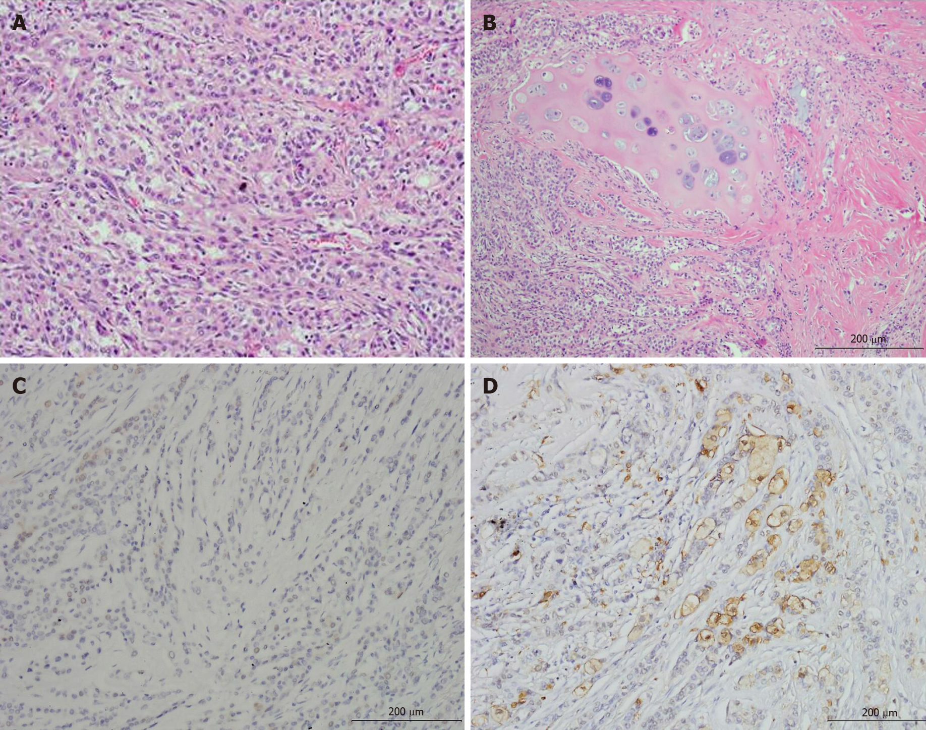
Figure 3 Pathology images.
A: Epithelial cells formed a duct-like structure, with occasional mucin production; B: Chondromyxoid and hyalinizing matrix as the background, and bronchial cartilage invasion were found; C: Weakly positive reactivity to synaptophysin in tumor cells indicated that a carcinoid lung tumor was unlikely; D: Myoepithelial cells positive for S-100 protein.
- Citation: Yang CH, Liu NT, Huang TW. Role of positron emission tomography in primary carcinoma ex pleomorphic adenoma of the bronchus: A case report. World J Clin Cases 2021; 9(12): 2811-2815
- URL: https://www.wjgnet.com/2307-8960/full/v9/i12/2811.htm
- DOI: https://dx.doi.org/10.12998/wjcc.v9.i12.2811


