Copyright
©The Author(s) 2021.
World J Clin Cases. Apr 26, 2021; 9(12): 2801-2810
Published online Apr 26, 2021. doi: 10.12998/wjcc.v9.i12.2801
Published online Apr 26, 2021. doi: 10.12998/wjcc.v9.i12.2801
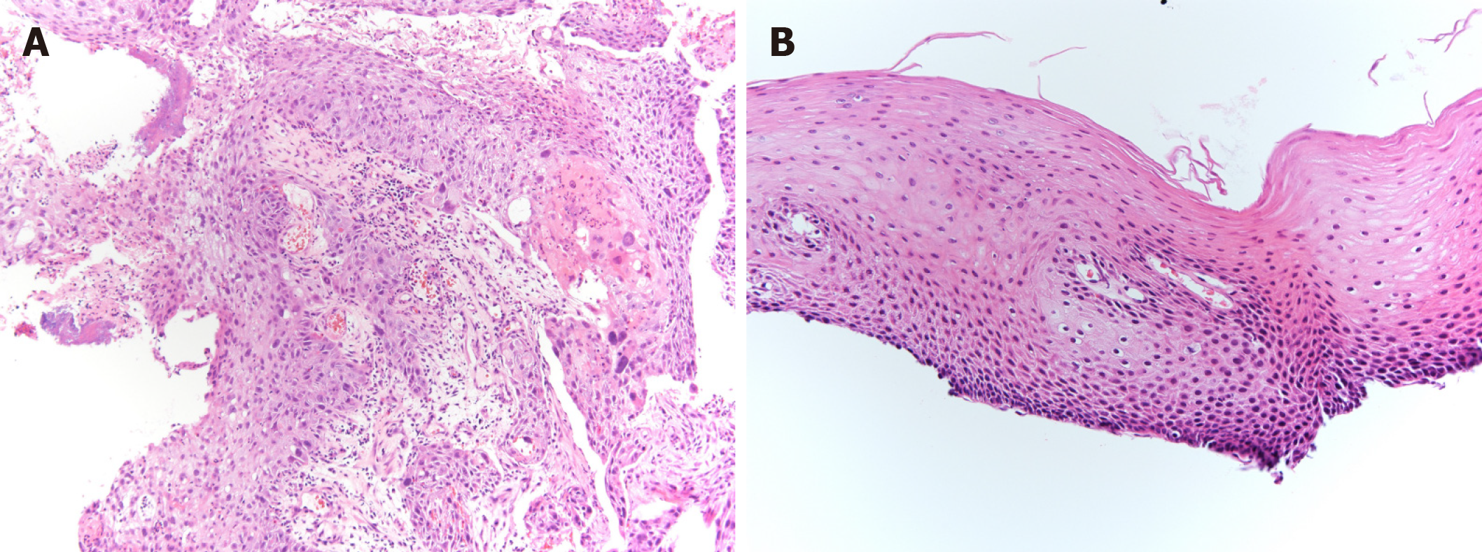
Figure 6 Pathological examination, biopsy from primary lesion.
A: Initial biopsy from primary tumor revealed squamous cell carcinoma (× 10). A proliferation of large atypical cells with swollen deep-stained nuclei was observed. A squamous cell carcinoma with a tendency toward keratinization, intercellular bridges, necrosis, and large atypical cells with bizarre nuclei were evident; B: Fourteen months after additional 2 courses of cisplatin plus 5fluorouracil chemotherapy, primary lesion has maintained pathologically negative for cancer (× 20). There is a small infiltration of inflammatory cells, but no obvious evidence of malignant cells.
- Citation: Yura M, Koyanagi K, Hara A, Hayashi K, Tajima Y, Kaneko Y, Fujisaki H, Hirata A, Takano K, Hongo K, Yo K, Yoneyama K, Tamai Y, Dehari R, Nakagawa M. Unresectable esophageal cancer treated with multiple chemotherapies in combination with chemoradiotherapy: A case report. World J Clin Cases 2021; 9(12): 2801-2810
- URL: https://www.wjgnet.com/2307-8960/full/v9/i12/2801.htm
- DOI: https://dx.doi.org/10.12998/wjcc.v9.i12.2801


