Copyright
©The Author(s) 2021.
World J Clin Cases. Apr 26, 2021; 9(12): 2791-2800
Published online Apr 26, 2021. doi: 10.12998/wjcc.v9.i12.2791
Published online Apr 26, 2021. doi: 10.12998/wjcc.v9.i12.2791
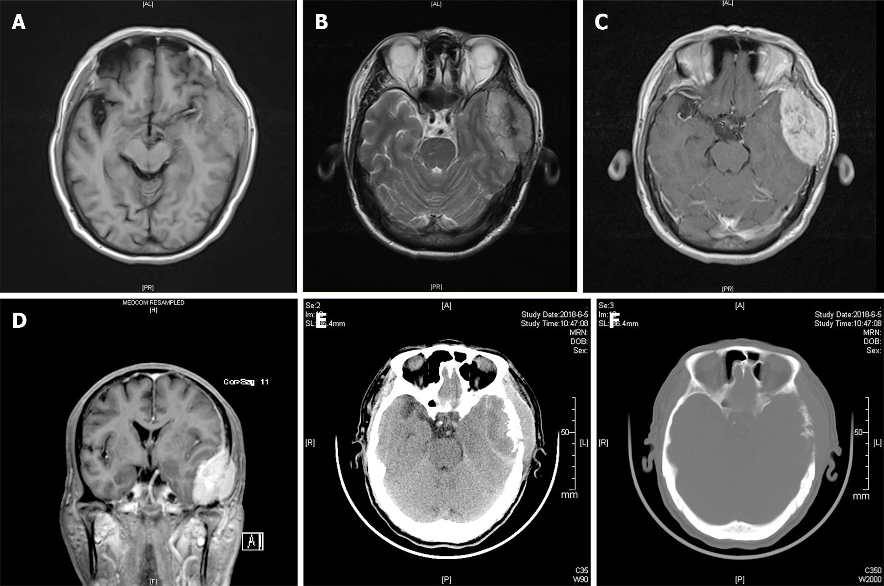
Figure 1 Radiographic images of the presenting case.
A: Magnetic resonance imaging (MRI) T1-weighted imaging showed an isointense left temporal lobe mass; B: MRI T2-weighted imaging showed a mixed hypointense and hyperintense left temporal lobe mass; C: Homogeneous enhancement of well-defined lesion was showed on axis MRI after intravenous contrast material; D: Coronal MRI showed the left temporal lobe mass after intravenous contrast material; E: Unenhanced computed tomography (CT) scan indicated the left temporal lobe mass; F: Multiple lytic lesion of the left temporal bone was noted on CT scan.
- Citation: Chen JC, Zhuang DZ, Luo C, Chen WQ. Malignant pheochromocytoma with cerebral and skull metastasis: A case report and literature review. World J Clin Cases 2021; 9(12): 2791-2800
- URL: https://www.wjgnet.com/2307-8960/full/v9/i12/2791.htm
- DOI: https://dx.doi.org/10.12998/wjcc.v9.i12.2791


