Copyright
©The Author(s) 2021.
World J Clin Cases. Apr 16, 2021; 9(11): 2634-2640
Published online Apr 16, 2021. doi: 10.12998/wjcc.v9.i11.2634
Published online Apr 16, 2021. doi: 10.12998/wjcc.v9.i11.2634
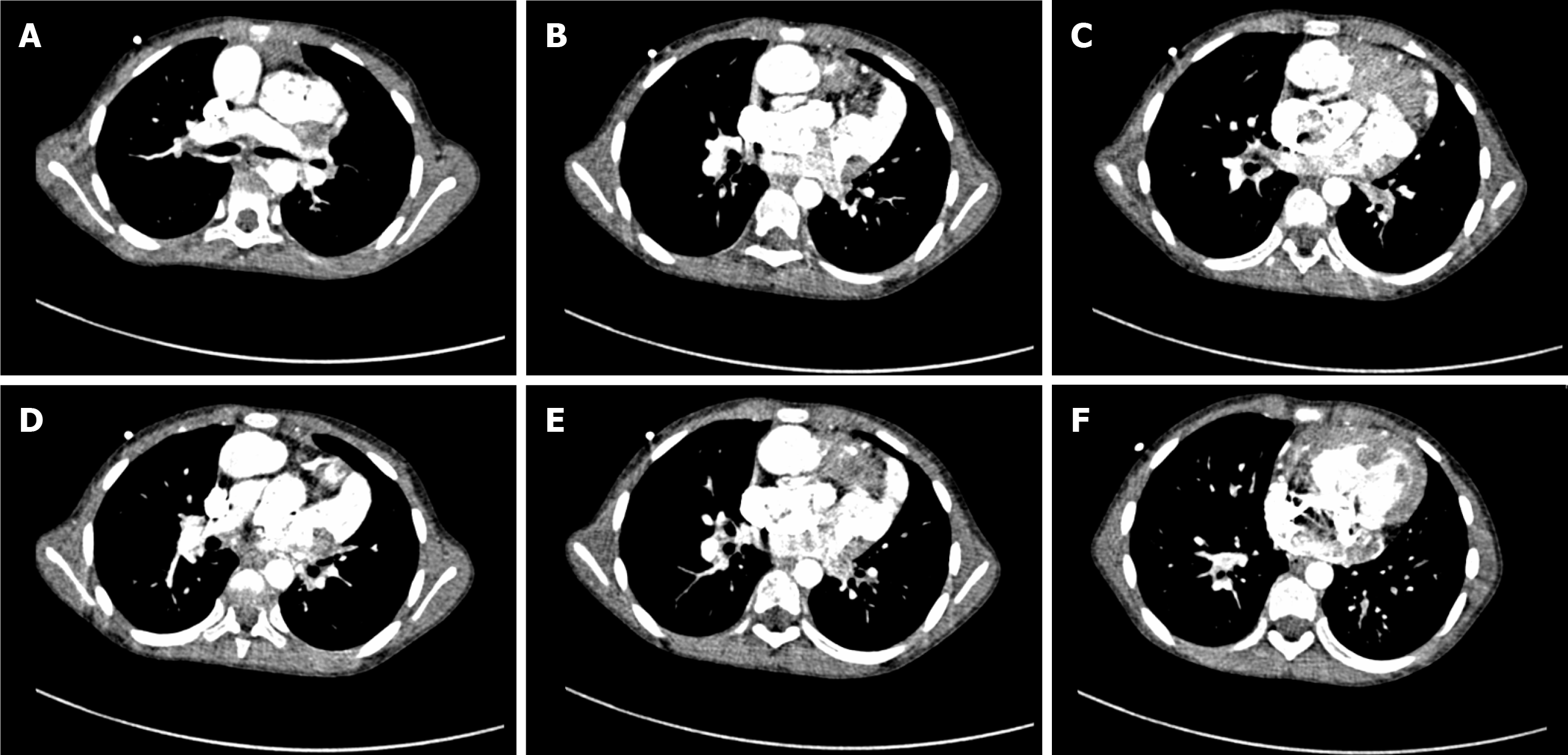
Figure 3 Chest computed tomography.
A-C: Both the aorta and pulmonary artery were connected to the right ventricle, and the aorta was anterior to the pulmonary artery; D-F: Significant right ventricular enlargement, atrial septal defect (width, 1.4 cm), and ventricular septal defect (width, 1.7 cm).
- Citation: Tan LC, Zhang WY, Zuo YD, Chen HY, Jiang CL. Anesthetic management of a child with double outlet right ventricle and severe polycythemia: A case report. World J Clin Cases 2021; 9(11): 2634-2640
- URL: https://www.wjgnet.com/2307-8960/full/v9/i11/2634.htm
- DOI: https://dx.doi.org/10.12998/wjcc.v9.i11.2634


