Copyright
©The Author(s) 2021.
World J Clin Cases. Apr 16, 2021; 9(11): 2542-2554
Published online Apr 16, 2021. doi: 10.12998/wjcc.v9.i11.2542
Published online Apr 16, 2021. doi: 10.12998/wjcc.v9.i11.2542
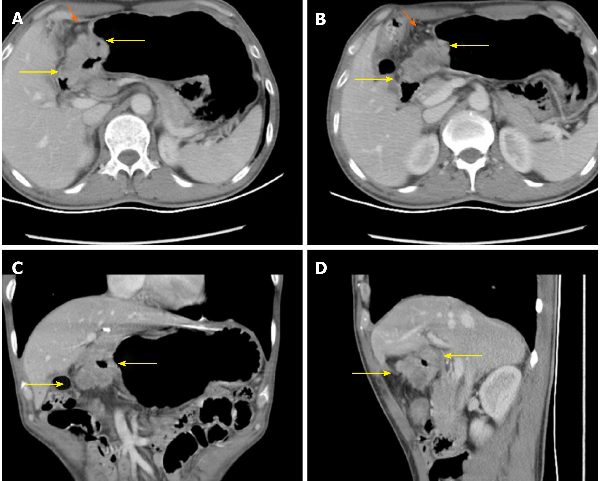
Figure 2 Enhanced abdominal computerized tomography indicated that the wall of the antrum was thickened with significant enhancement.
The surface of serosa was fuzzy, but the border near the pancreas was still clear (yellow arrowheads). Multiple enlarged lymph nodes were found in the lesser gastric curvature (orange arrowheads). A-B: Transverse views of the primary lesion in different layers; C: Coronal view of the primary lesion; D: Sagittal view of the primary lesion.
- Citation: Liu ZN, Wang YK, Li ZY. Neoadjuvant chemoradiotherapy followed by laparoscopic distal gastrectomy in advanced gastric cancer: A case report and review of literature. World J Clin Cases 2021; 9(11): 2542-2554
- URL: https://www.wjgnet.com/2307-8960/full/v9/i11/2542.htm
- DOI: https://dx.doi.org/10.12998/wjcc.v9.i11.2542


