Copyright
©The Author(s) 2021.
World J Clin Cases. Apr 16, 2021; 9(11): 2519-2523
Published online Apr 16, 2021. doi: 10.12998/wjcc.v9.i11.2519
Published online Apr 16, 2021. doi: 10.12998/wjcc.v9.i11.2519
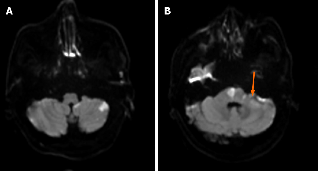
Figure 1 The results of diffusion-weighted imaging in patients with brain magnetic resonance imaging.
A: Diffusion-weighted imaging (DWI) revealed high signal intensity in the left posterior inferior cerebellar artery territory of the cerebellar hemisphere; B: DWI revealed high signal intensity in the right pons and bridge cerebellar arm. The yellow arrow indicates that the left auditory nerve was high signal on DWI, suggesting infarction of the auditory nerve.
- Citation: Wang XL, Sun M, Wang XP. Cerebellar artery infarction with sudden hearing loss and vertigo as initial symptoms: A case report. World J Clin Cases 2021; 9(11): 2519-2523
- URL: https://www.wjgnet.com/2307-8960/full/v9/i11/2519.htm
- DOI: https://dx.doi.org/10.12998/wjcc.v9.i11.2519


