Copyright
©The Author(s) 2021.
World J Clin Cases. Apr 6, 2021; 9(10): 2400-2408
Published online Apr 6, 2021. doi: 10.12998/wjcc.v9.i10.2400
Published online Apr 6, 2021. doi: 10.12998/wjcc.v9.i10.2400
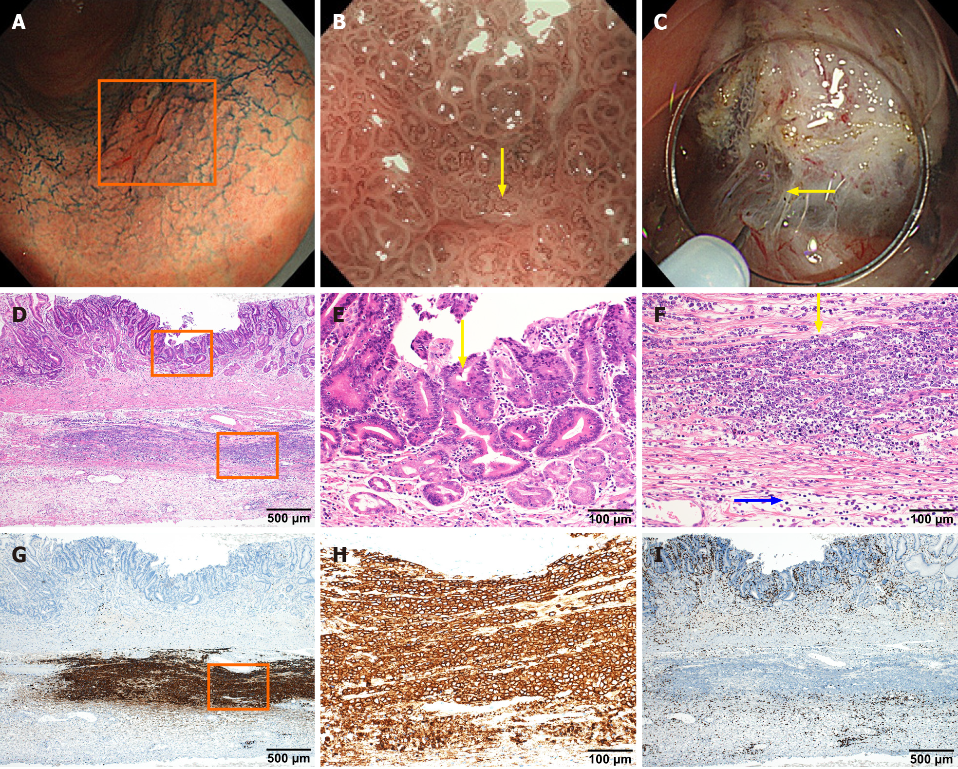
Figure 3 In situ collision between gastric cancer and diffuse large B-cell lymphoma.
A-C: Endoscopic images of lesions were obtained by electronic staining magnifying gastroscopy (GIF-H290Z). Indigo carmine local spray (0.2%) staining of mucous membrane shows uneven staining in the lesion area indicated by the box, with local redness, rough surface, but clear lesion border (A). Narrow band imaging at maximum 85 × magnification under GIF-H290Z gastroscopy clearly shows irregular villi-like structures on the surface of the lesion and vessels of different calibers as indicated by yellow arrows (B). The lesion fibrosis is obvious during endoscopic submucosal dissection operation, as indicated by the arrow, suggesting that the lesion is not confined to the mucosal layer (C); D: Histological examinations revealed that the resected gastric specimen contained a small ulcerated lesion (approximately 5 mm in length) composed of atypical epithelial cells with large nuclei; E: Tubular or papillary proliferation is present, but without submucosal invasion; F: Beneath the gastric carcinoma, there was diffuse infiltration of atypical lymphoid cells in the muscular propria. The yellow arrow indicates atypical lymphoid cells that are large in size, compared with the blue arrow that indicates normal lymphocytes; G-I: Immunohistochemical staining analysis, with brown staining showing abnormal lymphoid cells are positively stained with CD20 (G and H) but negatively stained with CD3 (I).
- Citation: Ma YH, Yamaguchi T, Yasumura T, Kuno T, Kobayashi S, Yoshida T, Ishida T, Ishida Y, Takaoka S, Fan JL, Enomoto N. Pancreatic cancer secondary to intraductal papillary mucinous neoplasm with collision between gastric cancer and B-cell lymphoma: A case report. World J Clin Cases 2021; 9(10): 2400-2408
- URL: https://www.wjgnet.com/2307-8960/full/v9/i10/2400.htm
- DOI: https://dx.doi.org/10.12998/wjcc.v9.i10.2400


