Copyright
©The Author(s) 2021.
World J Clin Cases. Apr 6, 2021; 9(10): 2380-2385
Published online Apr 6, 2021. doi: 10.12998/wjcc.v9.i10.2380
Published online Apr 6, 2021. doi: 10.12998/wjcc.v9.i10.2380
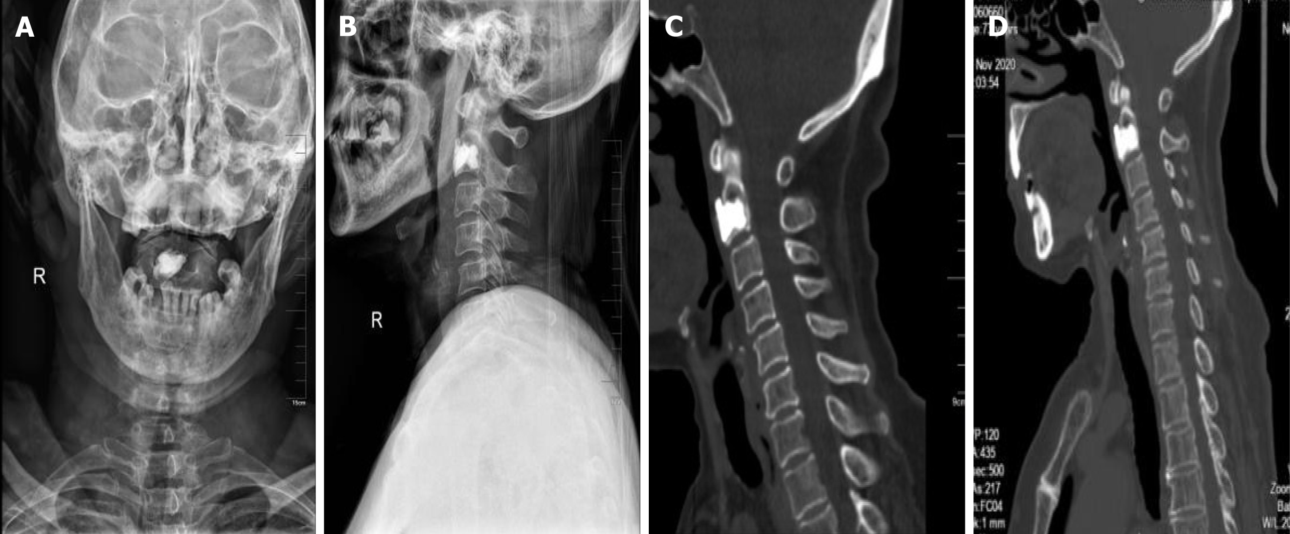
Figure 5 Postoperative X-rays and computed tomography showed that the bone cement filled well, and no leakage occurred during 3 years of follow-up.
A and B: The anteroposterior and lateral X-ray was taken at 2 d after surgery; C and D: The two sagittal computed tomography scans were taken at 6 mo and 3 yr after surgery. They showed that no leakage of the bone cement occurred, and the shape of the C2 was maintained.
- Citation: Li RJ, Li XF, Jiang WM. Solitary bone plasmacytoma of the upper cervical spine: A case report . World J Clin Cases 2021; 9(10): 2380-2385
- URL: https://www.wjgnet.com/2307-8960/full/v9/i10/2380.htm
- DOI: https://dx.doi.org/10.12998/wjcc.v9.i10.2380


