Copyright
©The Author(s) 2020.
World J Clin Cases. Jan 6, 2021; 9(1): 61-70
Published online Jan 6, 2021. doi: 10.12998/wjcc.v9.i1.61
Published online Jan 6, 2021. doi: 10.12998/wjcc.v9.i1.61
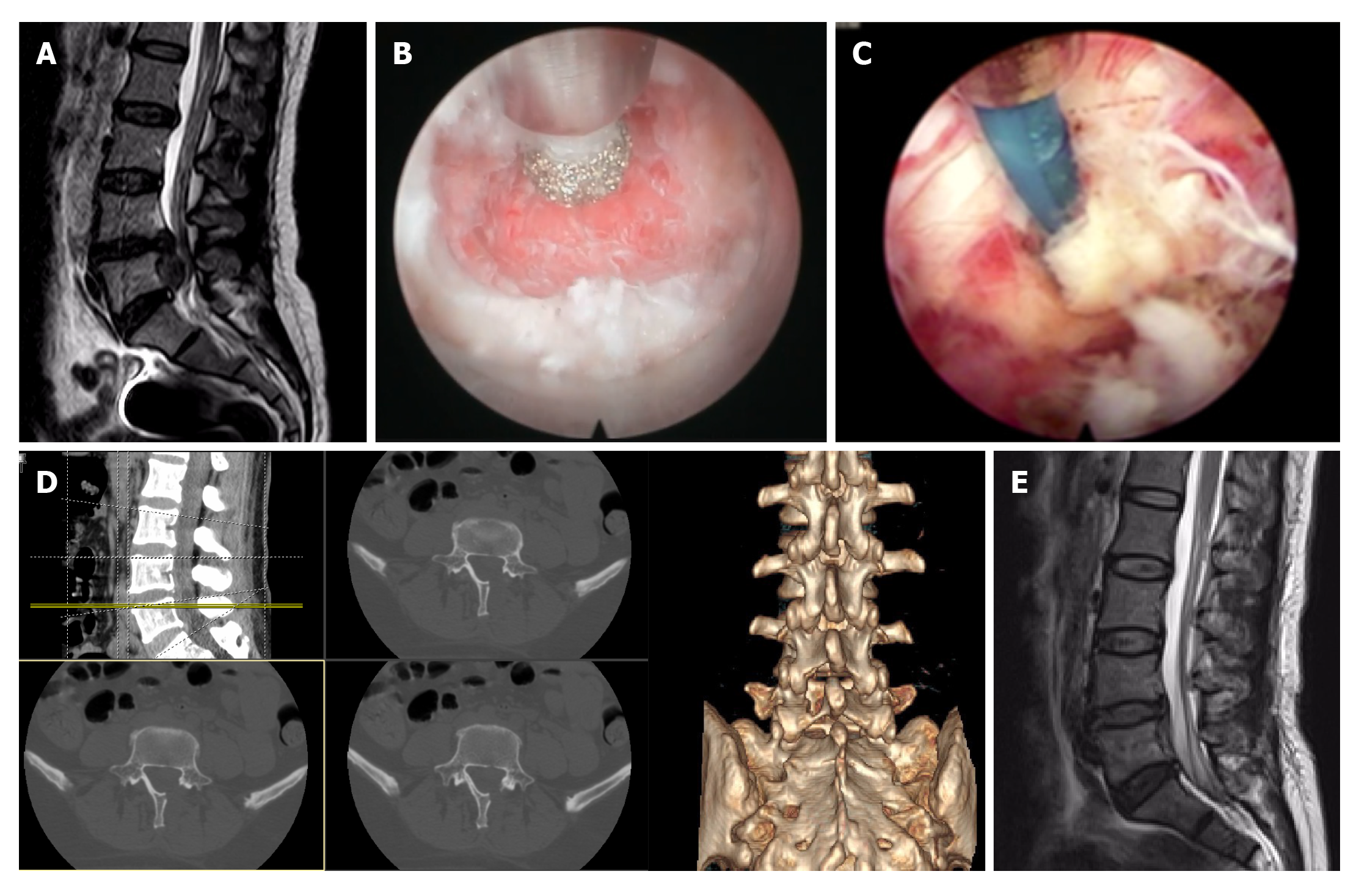
Figure 2 An endoscopic bur and bone ribbing rongeur for vertebral plate was used to remove a part of the vertebral plate and increase the laminae interval space, while the zygopophysis was protected.
A: Preoperative sagittal magnetic resonance imaging (MRI) showing a far inferiorly migrated L4/L5 Lumbar disc herniation, of which the distal end was close to the inferior endplate of L5, and the spinal sac was severely compressed; B: Use of an endoscopic bur to enlarge the interlaminar space; C: Searching for the nerve roots and protruding vertebral pulp under the endoscopy; D: Postoperative re-examination with computed tomography showing the size and range of the abraded vertebral plate; E: Postoperative MRI showing no residual vertebral pulp in the case of inferiorly migrated L4/L5 lumbar disc herniation, and there was no evident compression on the spinal sac.
- Citation: Meng SW, Peng C, Zhou CL, Tao H, Wang C, Zhu K, Song MX, Ma XX. Massively prolapsed intervertebral disc herniation with interlaminar endoscopic spine system Delta endoscope: A case series. World J Clin Cases 2021; 9(1): 61-70
- URL: https://www.wjgnet.com/2307-8960/full/v9/i1/61.htm
- DOI: https://dx.doi.org/10.12998/wjcc.v9.i1.61


