Copyright
©The Author(s) 2021.
World J Clin Cases. Jan 6, 2021; 9(1): 278-283
Published online Jan 6, 2021. doi: 10.12998/wjcc.v9.i1.278
Published online Jan 6, 2021. doi: 10.12998/wjcc.v9.i1.278
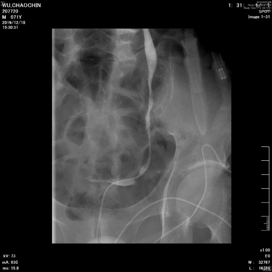
Figure 3 Left ureteral stenosis.
Left retrograde pyelography showed multiple stenosis and narrowing points along middle to lower ureter, which led to left hydronephrosis and hydroureter.
- Citation: Tsao SH, Chuang CK. Krukenberg tumor with concomitant ipsilateral hydronephrosis and spermatic cord metastasis in a man: A case report. World J Clin Cases 2021; 9(1): 278-283
- URL: https://www.wjgnet.com/2307-8960/full/v9/i1/278.htm
- DOI: https://dx.doi.org/10.12998/wjcc.v9.i1.278


