Copyright
©The Author(s) 2021.
World J Clin Cases. Jan 6, 2021; 9(1): 218-223
Published online Jan 6, 2021. doi: 10.12998/wjcc.v9.i1.218
Published online Jan 6, 2021. doi: 10.12998/wjcc.v9.i1.218
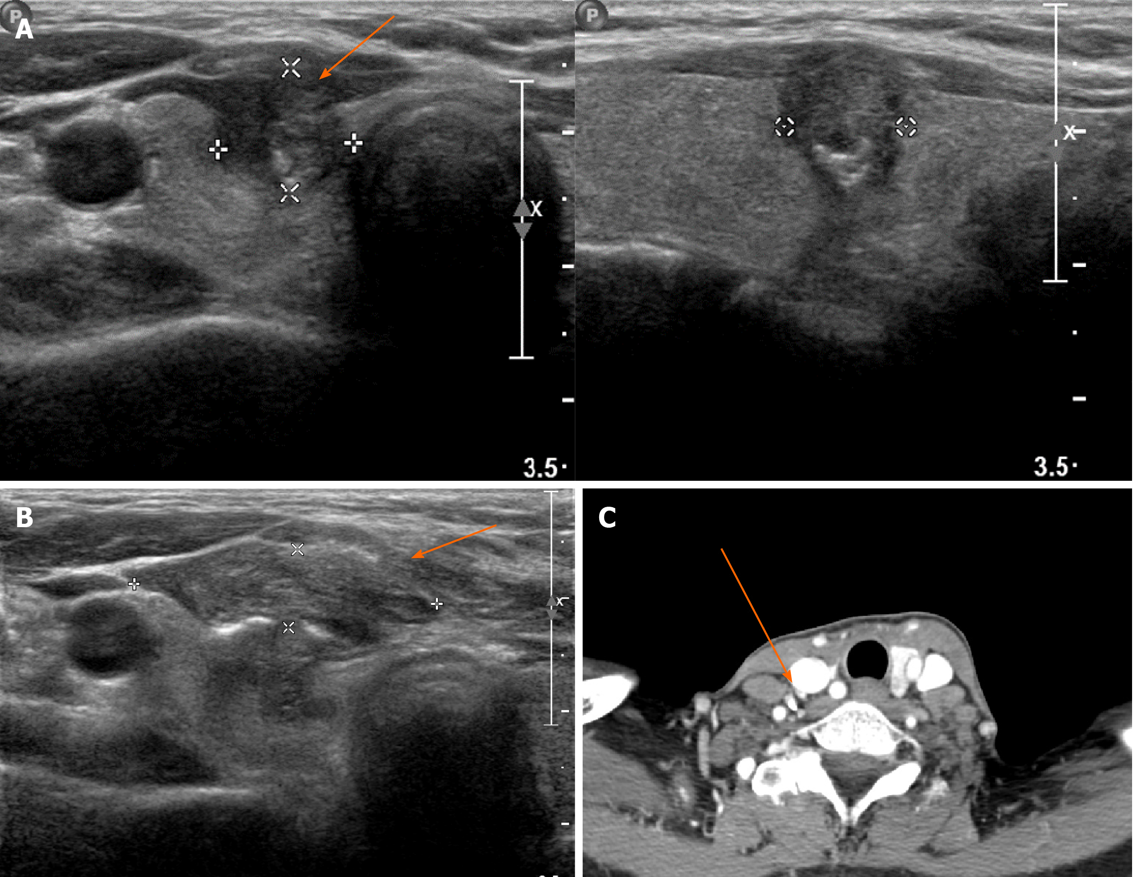
Figure 1 Radiologic images of initial diagnosis of papillary thyroid cancer.
A: Tumor located in right middle lobe of the thyroid; B: Postprocedural hematoma after core needle biopsy through isthmus observed by ultrasonography; C: Presents computed tomography of enlarged lymph node at level IV, performed after excisional biopsy of skin tumor.
- Citation: Kim YH, Choi IH, Lee JE, Kim Z, Han SW, Hur SM, Lee J. Late recurrence of papillary thyroid cancer from needle tract implantation after core needle biopsy: A case report. World J Clin Cases 2021; 9(1): 218-223
- URL: https://www.wjgnet.com/2307-8960/full/v9/i1/218.htm
- DOI: https://dx.doi.org/10.12998/wjcc.v9.i1.218


