Copyright
©The Author(s) 2020.
World J Clin Cases. May 6, 2020; 8(9): 1721-1728
Published online May 6, 2020. doi: 10.12998/wjcc.v8.i9.1721
Published online May 6, 2020. doi: 10.12998/wjcc.v8.i9.1721
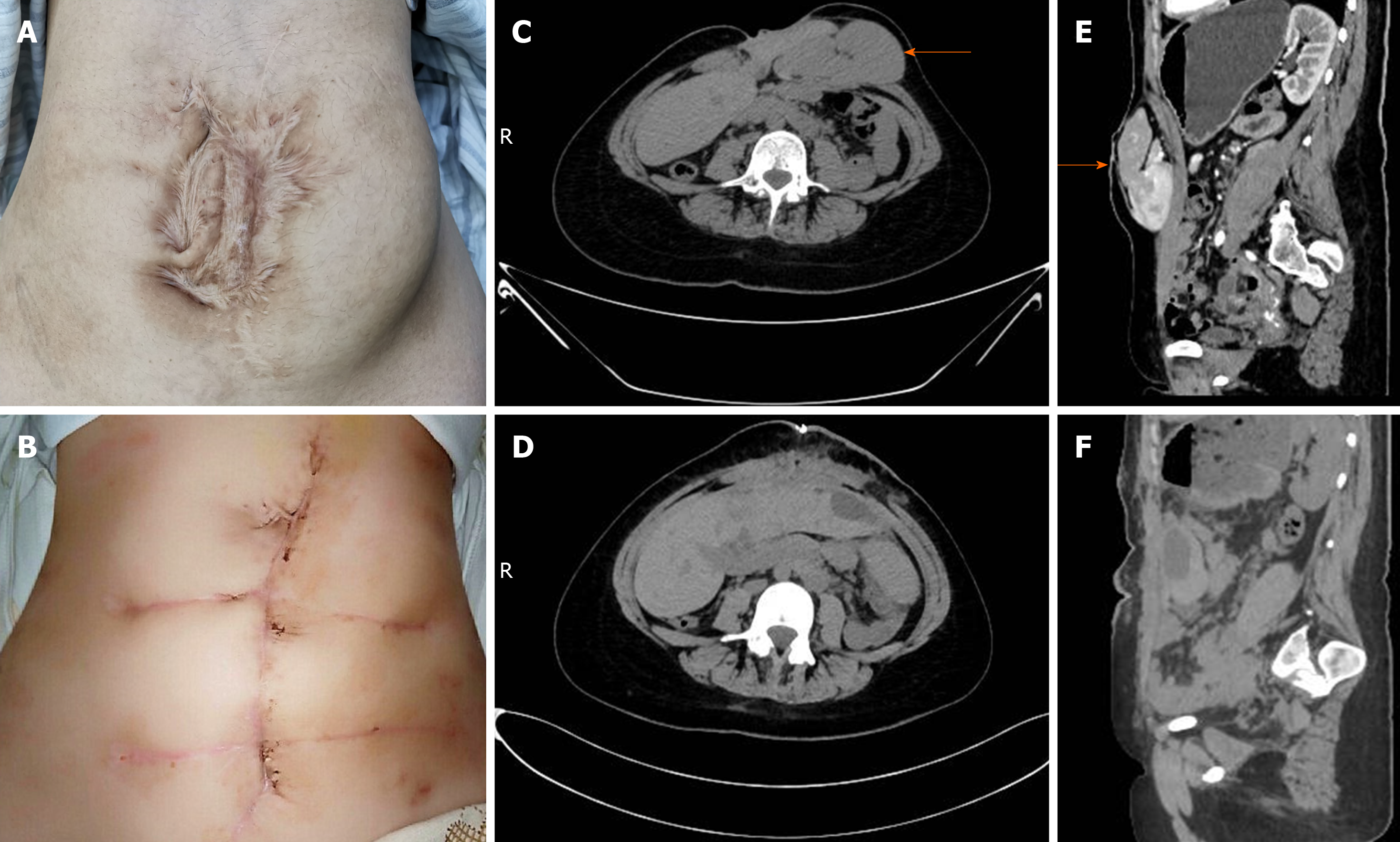
Figure 1 Patient-related image.
A: Many irregular and obsolete surgical scars on the abdominal wall; B: The appearance of the surgical incision improved greatly after operation; C and E: Computed tomography demonstrated a giant ventral hernia containing liver, pancreas, spleen and blood vessels before operation (arrow); D and F: Liver, pancreas, spleen and blood vessels returned to the abdominal cavity after operation.
- Citation: Luo XG, Lu C, Wang WL, Zhou F, Yu CZ. Giant ventral hernia simultaneously containing the spleen, a portion of the pancreas and the left hepatic lobe: A case report. World J Clin Cases 2020; 8(9): 1721-1728
- URL: https://www.wjgnet.com/2307-8960/full/v8/i9/1721.htm
- DOI: https://dx.doi.org/10.12998/wjcc.v8.i9.1721


