Copyright
©The Author(s) 2020.
World J Clin Cases. May 6, 2020; 8(9): 1698-1704
Published online May 6, 2020. doi: 10.12998/wjcc.v8.i9.1698
Published online May 6, 2020. doi: 10.12998/wjcc.v8.i9.1698
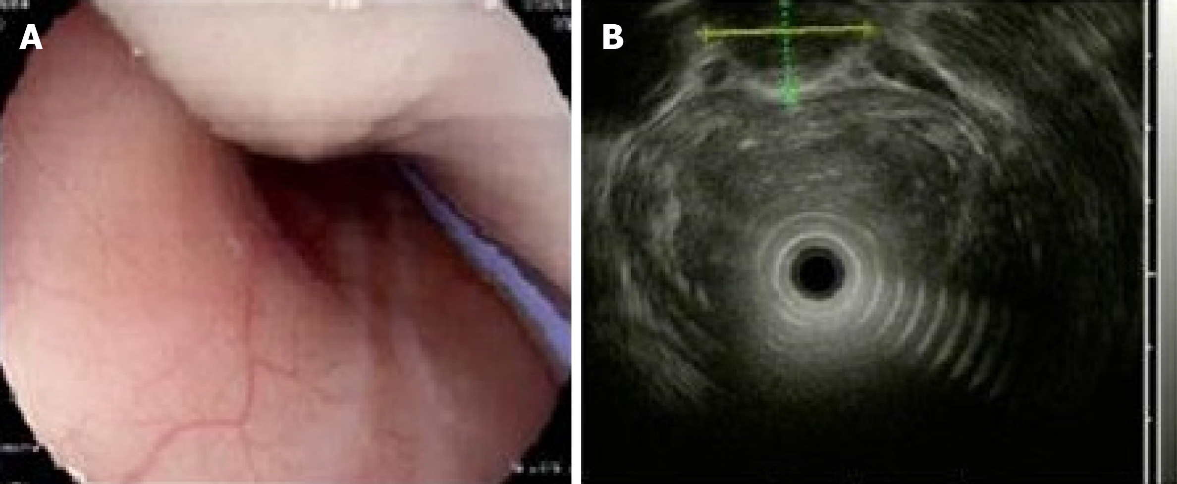
Figure 2 Esophagogastric ultrasonography showed a homogeneous, low echogenic mass outside the esophagus.
A: Esophagogastroscopy showed a large mass pushing against the esophagus; B: Esophagogastric ultrasonography showed a large, homogeneous, low echogenic mass outside the esophagus, 20–40 cm below the incisor, with a cross-sectional area of 21.5 mm × 15.9 mm.
- Citation: Ye YW, Liao MY, Mou ZM, Shi XX, Xie YC. Thoracoscopic resection of a huge esophageal dedifferentiated liposarcoma: A case report. World J Clin Cases 2020; 8(9): 1698-1704
- URL: https://www.wjgnet.com/2307-8960/full/v8/i9/1698.htm
- DOI: https://dx.doi.org/10.12998/wjcc.v8.i9.1698


