Copyright
©The Author(s) 2020.
World J Clin Cases. Apr 26, 2020; 8(8): 1525-1531
Published online Apr 26, 2020. doi: 10.12998/wjcc.v8.i8.1525
Published online Apr 26, 2020. doi: 10.12998/wjcc.v8.i8.1525
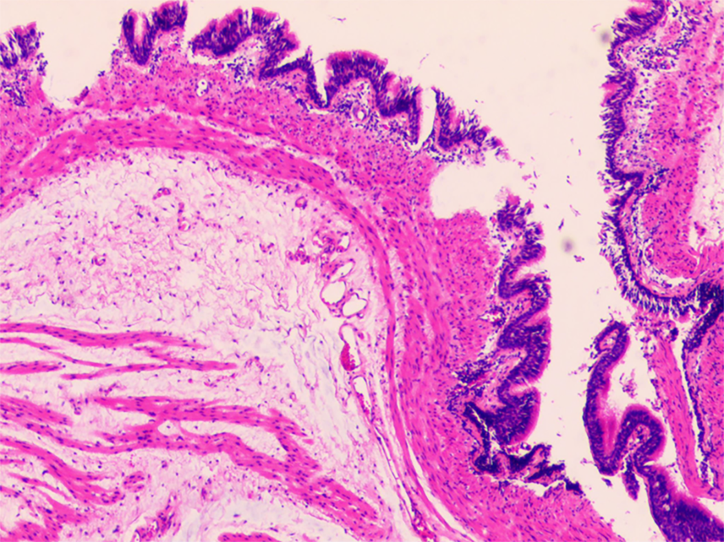
Figure 3 Postoperative histopathology.
Microscopically, pseudostratified ciliated columnar epithelial cells could be observed in the cyst wall, and the structure of these cells was the same as that of the bronchus. Combined with immunohistochemical staining for CK7 (+), CK20 (-), TTF1 (-), and S-100 (-), the pathologists ultimately verified that this cystic mass of the fundus was a gastric bronchogenic cyst (×100).
- Citation: He WT, Deng JY, Liang H, Xiao JY, Cao FL. Bronchogenic cyst of the stomach: A case report. World J Clin Cases 2020; 8(8): 1525-1531
- URL: https://www.wjgnet.com/2307-8960/full/v8/i8/1525.htm
- DOI: https://dx.doi.org/10.12998/wjcc.v8.i8.1525


