Copyright
©The Author(s) 2020.
World J Clin Cases. Apr 26, 2020; 8(8): 1525-1531
Published online Apr 26, 2020. doi: 10.12998/wjcc.v8.i8.1525
Published online Apr 26, 2020. doi: 10.12998/wjcc.v8.i8.1525
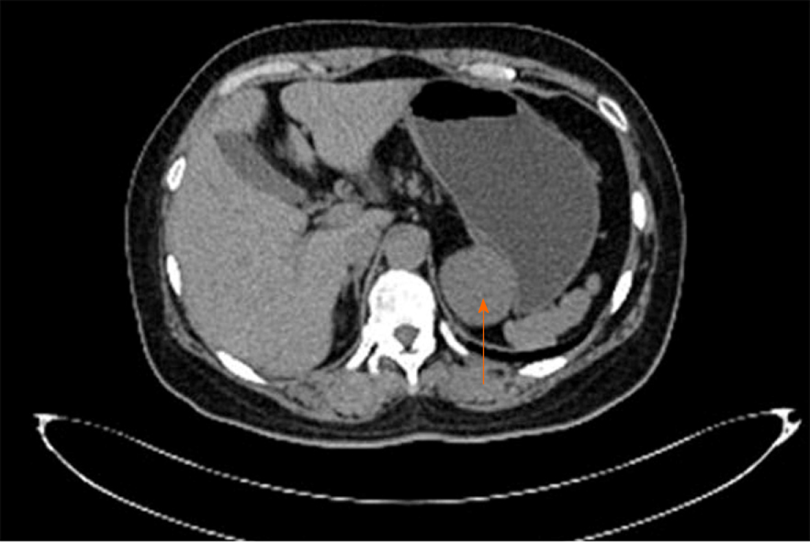
Figure 2 Imaging at admission.
A plain computed tomography scan demonstrated a 52 mm × 43 mm quasi-circular mass (arrow). It appeared to have a low density of 29 HU and was closely related to the lesser curvature of the gastric fundus, which also showed an extraluminal growth pattern with an obvious border.
- Citation: He WT, Deng JY, Liang H, Xiao JY, Cao FL. Bronchogenic cyst of the stomach: A case report. World J Clin Cases 2020; 8(8): 1525-1531
- URL: https://www.wjgnet.com/2307-8960/full/v8/i8/1525.htm
- DOI: https://dx.doi.org/10.12998/wjcc.v8.i8.1525


