Copyright
©The Author(s) 2020.
World J Clin Cases. Apr 26, 2020; 8(8): 1525-1531
Published online Apr 26, 2020. doi: 10.12998/wjcc.v8.i8.1525
Published online Apr 26, 2020. doi: 10.12998/wjcc.v8.i8.1525
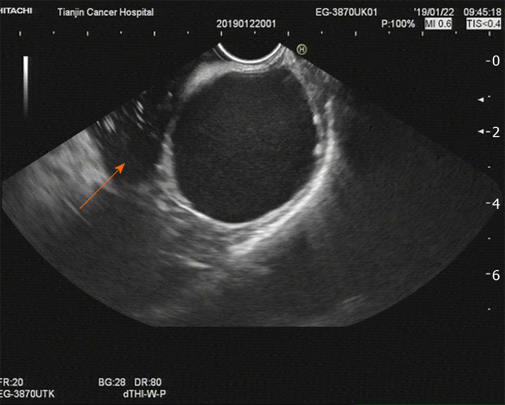
Figure 1 Imaging at admission.
An endoscopic ultrasound examination showed a 45 mm × 49 mm single cyst originating from the gastric submucosa (arrow). It also revealed that the cyst, without any echoes or color flow signals, was located in the posterior wall of the fundus close to the cardia. These results indicated the possibility of cystic hygroma of the stomach.
- Citation: He WT, Deng JY, Liang H, Xiao JY, Cao FL. Bronchogenic cyst of the stomach: A case report. World J Clin Cases 2020; 8(8): 1525-1531
- URL: https://www.wjgnet.com/2307-8960/full/v8/i8/1525.htm
- DOI: https://dx.doi.org/10.12998/wjcc.v8.i8.1525


