
















©The Author(s) 2020.
World J Clin Cases. Apr 26, 2020; 8(8): 1515-1524
Published online Apr 26, 2020. doi: 10.12998/wjcc.v8.i8.1515
Published online Apr 26, 2020. doi: 10.12998/wjcc.v8.i8.1515
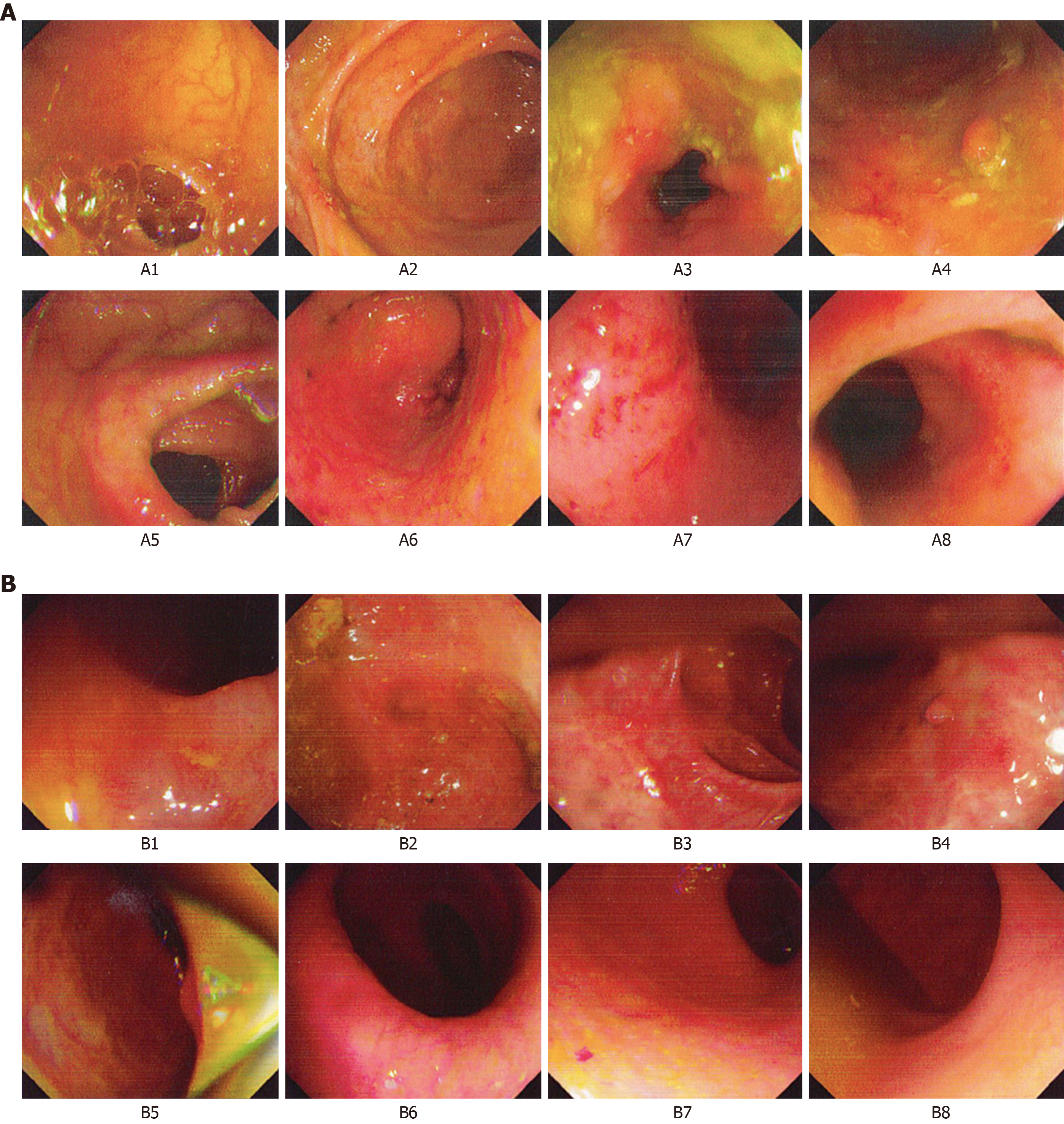
Figure 1 Comparison of colonoscopy results before and after treatment.
A: Before treatment (March 13, 2018); B: After treatment (January 10, 2019). A1: End of ileum; A2: Ileocecum; A3: Ascending colon; A4: Hepatic flexure; A5: Transverse colon; A6: Descending colon; A7: Rectum; A8: Anus; under endoscope, mucosal edema and hyperemia were observed in A3 (ascending colon), A4 (hepatic flexure), A6 (descending colon), sigmoid colon, and A7 (rectum), which showed active ulcerative colitis. B1: End of ileum; B2: Ileocecum; B3: Ascending colon; B4: Transverse colon; B5: Descending colon; B6: Sigmoid colon; B7: Rectum; and B8: Anus; under endoscopy, ulcer scars and inflammatory polyps were visible in part of the intestines, no ulcer or abnormal protuberance was observed, and the mucosa was nearly healed.
- Citation: Lin YY, Zhao JM, Ji YJ, Ma Z, Zheng HD, Huang Y, Cui YH, Lu Y, Wu HG. Typical ulcerative colitis treated by herbs-partitioned moxibustion: A case report. World J Clin Cases 2020; 8(8): 1515-1524
- URL: https://www.wjgnet.com/2307-8960/full/v8/i8/1515.htm
- DOI: https://dx.doi.org/10.12998/wjcc.v8.i8.1515