Copyright
©The Author(s) 2020.
World J Clin Cases. Feb 26, 2020; 8(4): 782-789
Published online Feb 26, 2020. doi: 10.12998/wjcc.v8.i4.782
Published online Feb 26, 2020. doi: 10.12998/wjcc.v8.i4.782
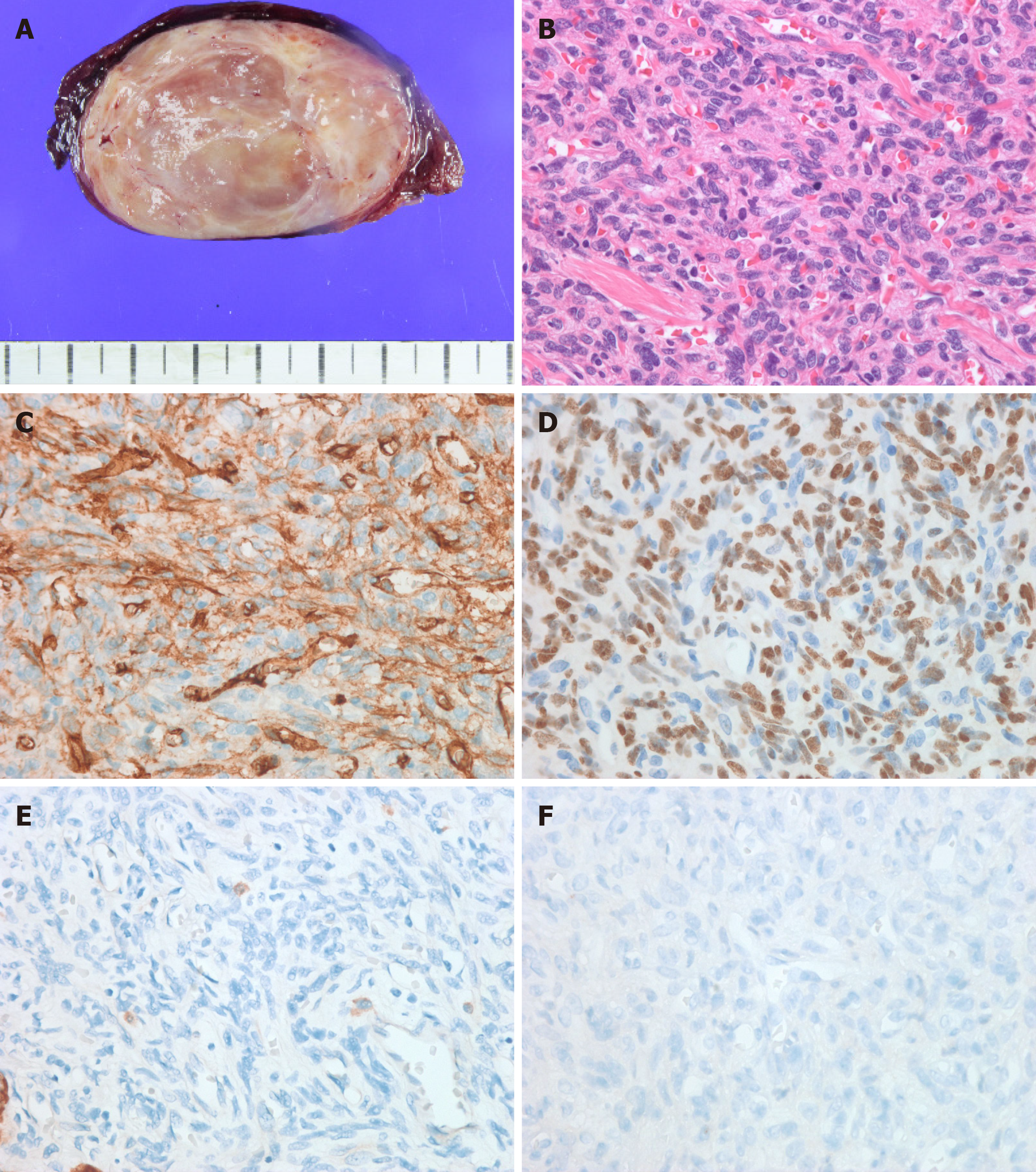
Figure 3 istology of a surgical specimen examined under light microscope.
A: Gross image; B: Hematoxylin and eosin staining (LM, × 40); C: CD34 (LM, × 400); D: Signal transducer and activator of transcription 6 (LM, × 400); E: CK (LM, × 400); F: TTF-1 (LM, × 400). LM: Light microscope.
- Citation: Suh YJ, Park JH, Jeon JH, Bilegsaikhan SE. Extrapleural solitary fibrous tumor of the thyroid gland: A case report and review of literature. World J Clin Cases 2020; 8(4): 782-789
- URL: https://www.wjgnet.com/2307-8960/full/v8/i4/782.htm
- DOI: https://dx.doi.org/10.12998/wjcc.v8.i4.782


