Copyright
©The Author(s) 2020.
World J Clin Cases. Feb 6, 2020; 8(3): 577-586
Published online Feb 6, 2020. doi: 10.12998/wjcc.v8.i3.577
Published online Feb 6, 2020. doi: 10.12998/wjcc.v8.i3.577
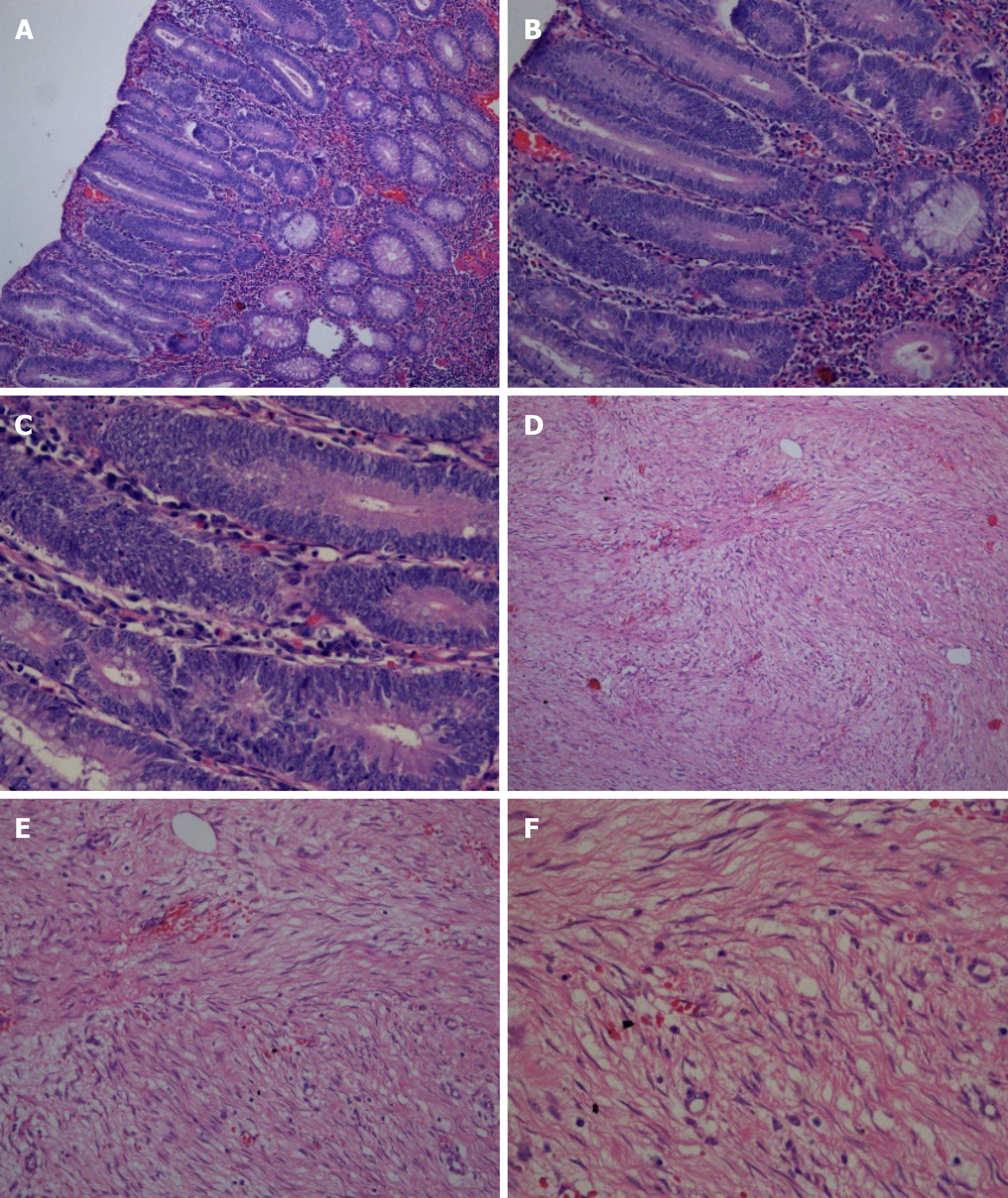
Figure 4 Pathological images of the patient.
A-C: Pathological images showed multiple tubularvillous adenomas with moderate dysplasia [A: hematoxylin and eosin (H&E) staining, 100 ×, B: H&E staining, 200 × and C: H&E staining, 400 ×]; D-F: Fusiform cell tumor-like hyperplasia of the mesentery (D: H&E staining, 100 ×, E: H&E staining, 200 × and F: H&E staining, 400 ×).
- Citation: Cai HJ, Wang H, Cao N, Wang W, Sun XX, Huang B. Peutz-Jeghers syndrome with mesenteric fibromatosis: A case report and review of literature. World J Clin Cases 2020; 8(3): 577-586
- URL: https://www.wjgnet.com/2307-8960/full/v8/i3/577.htm
- DOI: https://dx.doi.org/10.12998/wjcc.v8.i3.577


