Copyright
©The Author(s) 2020.
World J Clin Cases. Dec 26, 2020; 8(24): 6487-6498
Published online Dec 26, 2020. doi: 10.12998/wjcc.v8.i24.6487
Published online Dec 26, 2020. doi: 10.12998/wjcc.v8.i24.6487
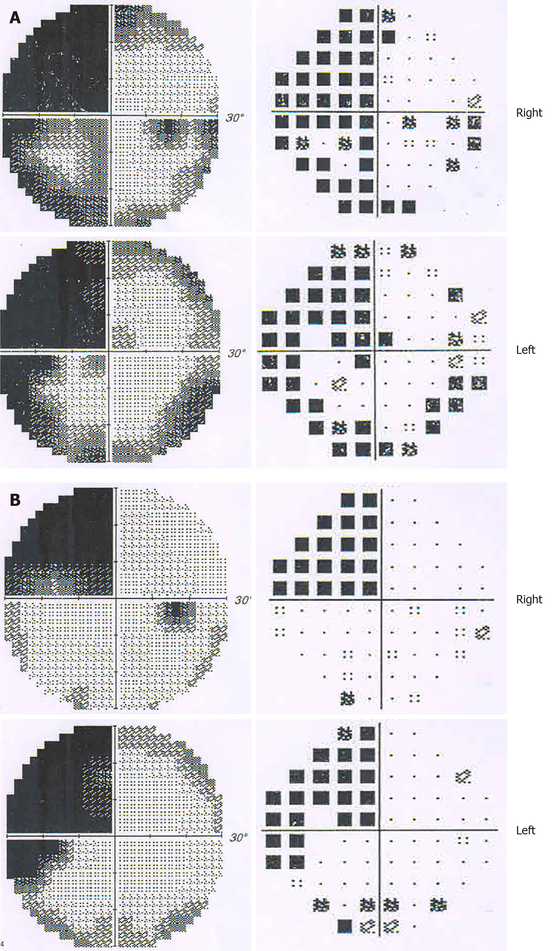
Figure 2 Visual field defect test showing homonymous left upper quadrantanopia.
A: Results of greyscale and pattern deviation at the onset of the stroke. Visual field index (VFI) was 56% in the right eye, and 61% in the left; B: Results of greyscale and pattern deviation after one month. VFI was 74% in both eyes.
- Citation: Yuan Y, Huang F, Gao ZH, Cai WC, Xiao JX, Yang YE, Zhu PL. Delayed diagnosis of prosopagnosia following a hemorrhagic stroke in an elderly man: A case report. World J Clin Cases 2020; 8(24): 6487-6498
- URL: https://www.wjgnet.com/2307-8960/full/v8/i24/6487.htm
- DOI: https://dx.doi.org/10.12998/wjcc.v8.i24.6487


