Copyright
©The Author(s) 2020.
World J Clin Cases. Dec 26, 2020; 8(24): 6425-6431
Published online Dec 26, 2020. doi: 10.12998/wjcc.v8.i24.6425
Published online Dec 26, 2020. doi: 10.12998/wjcc.v8.i24.6425
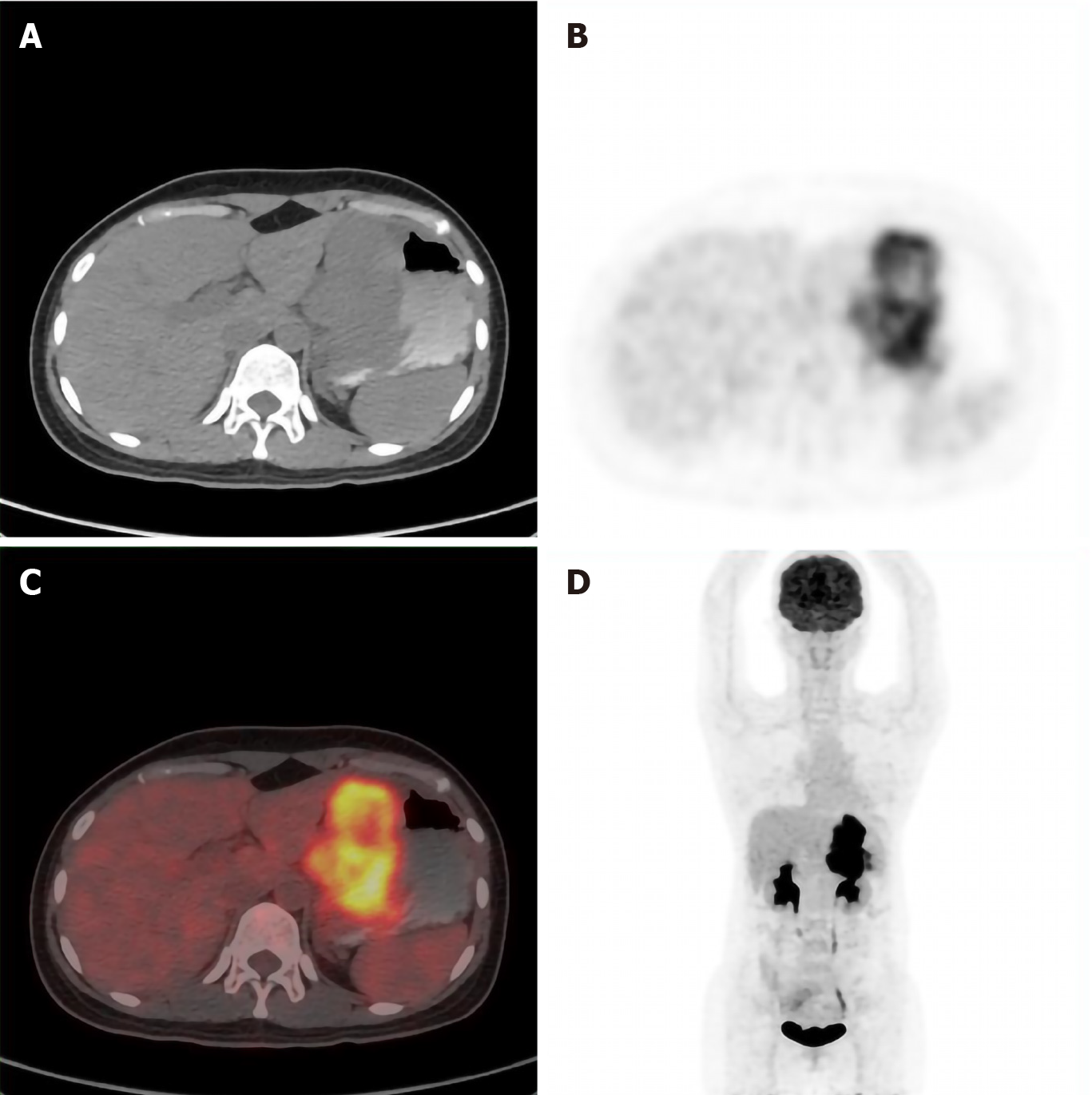
Figure 3 Whole body positron emission tomography-computed tomography showing tumor in the stomach only, with no other primary lesions.
A: Plain computed tomography (CT) scan of the abdomen; B: Abdominal positron emission tomography (PET)-CT; C: Fusion diagram of the scan shown in A and B. Color overlay shows the tracer uptake; D: Whole body coronal PET-CT. The gastric wall was markedly thickened and the glucose metabolism to be substantially increased. A large amount of metabolic radioactive concentration was seen in the bladder.
- Citation: Long GJ, Ou WT, Lin L, Zhou CJ. Primary gastric melanoma in a young woman: A case report. World J Clin Cases 2020; 8(24): 6425-6431
- URL: https://www.wjgnet.com/2307-8960/full/v8/i24/6425.htm
- DOI: https://dx.doi.org/10.12998/wjcc.v8.i24.6425


