Copyright
©The Author(s) 2020.
World J Clin Cases. Dec 26, 2020; 8(24): 6408-6417
Published online Dec 26, 2020. doi: 10.12998/wjcc.v8.i24.6408
Published online Dec 26, 2020. doi: 10.12998/wjcc.v8.i24.6408
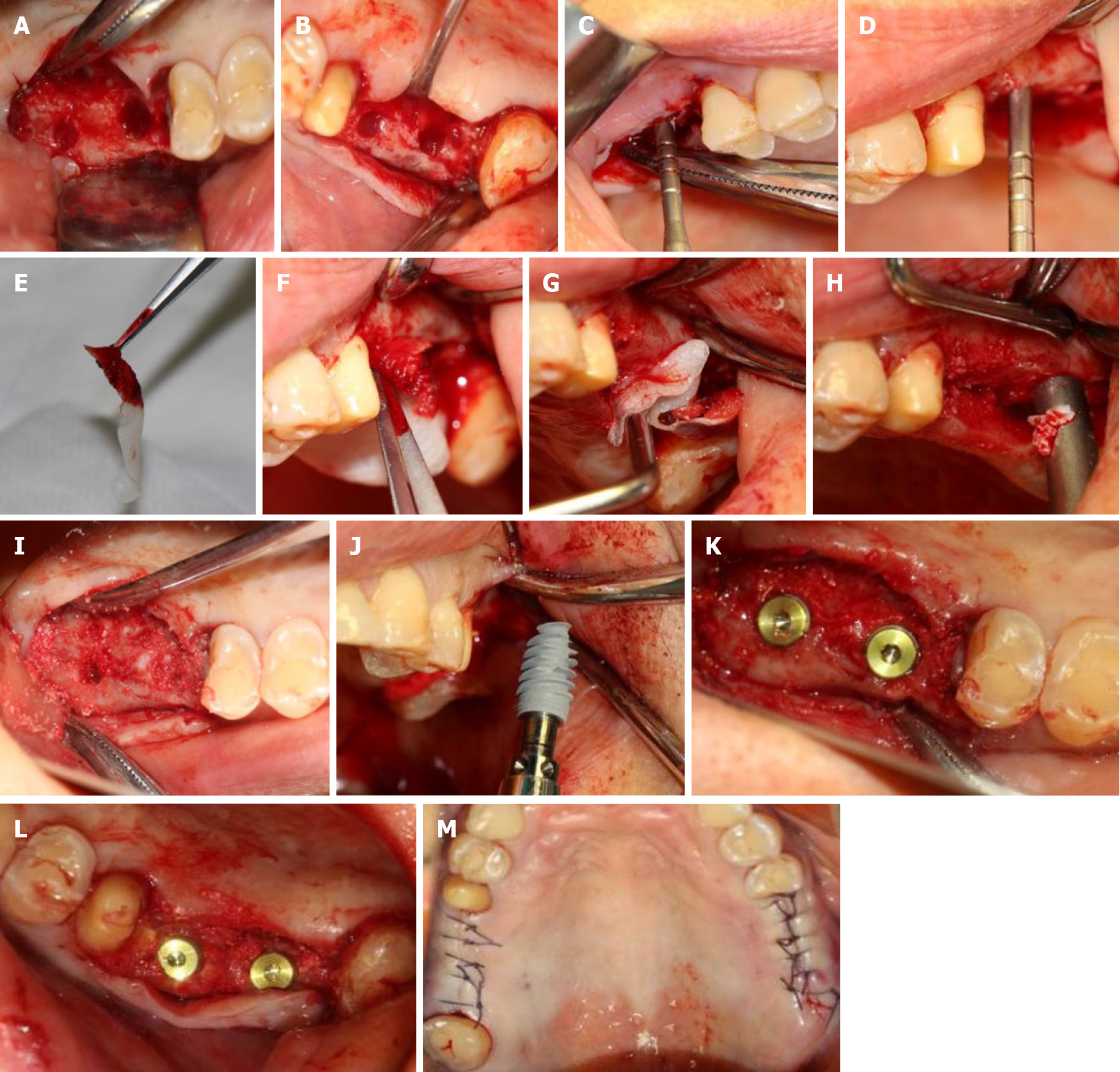
Figure 2 First-stage surgery.
A and B: Nest preparation on the right; C and D: Bilateral internal maxillary sinus floor elevation; E: Platelet-rich fibrin (PRF) clots were compressed between sterile dry gauze; F and G: Established PRF membranes were placed in the primary elevated sinus floor; H and I: Implantation of the bovine bone graft material; J: Implant (4.3 mm × 10 mm, Nobel Active, Sweden) was implanted with a torque of 35 N·cm; K and L: Placement of a suitable cover screw; M: Stitches in the wound.
- Citation: Yang S, Yu W, Zhang J, Zhou Z, Meng F, Wang J, Shi R, Zhou YM, Zhao J. Minimally invasive maxillary sinus augmentation with simultaneous implantation on an elderly patient: A case report. World J Clin Cases 2020; 8(24): 6408-6417
- URL: https://www.wjgnet.com/2307-8960/full/v8/i24/6408.htm
- DOI: https://dx.doi.org/10.12998/wjcc.v8.i24.6408


