Copyright
©The Author(s) 2020.
World J Clin Cases. Dec 26, 2020; 8(24): 6364-6372
Published online Dec 26, 2020. doi: 10.12998/wjcc.v8.i24.6364
Published online Dec 26, 2020. doi: 10.12998/wjcc.v8.i24.6364
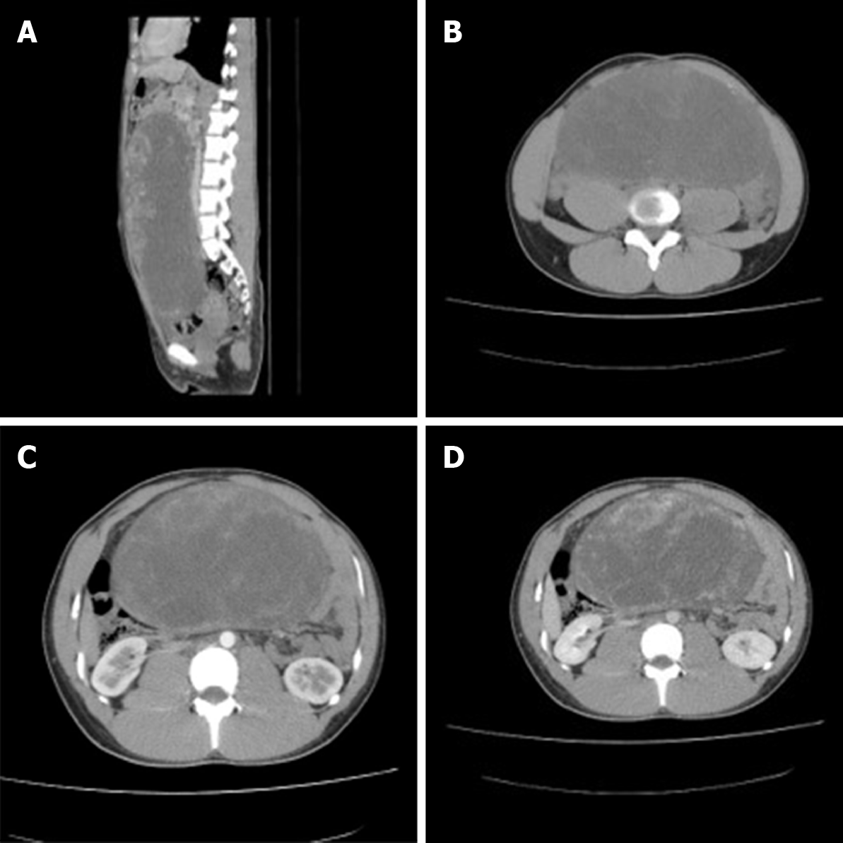
Figure 2 Computed tomography scan of the tumor.
A: Non-enhanced computed tomography (CT) scan showing a large, solid and cystic, partly calcified pelvic mass of the right ovary that measured 27.1 cm × 20.0 cm × 11.0 cm; B: The tumor had clear boundaries and showed non-homogeneous density with solid tissue at the periphery; C: Dynamic contrast-enhanced CT scan showing early peripheral ring enhancement associated with patch enhancement in the center of the lesion; D: In the venous phase, the tumor demonstrated progressive, centripetal and prolonged enhancement, the central portion of the tumor was not enhanced.
- Citation: Chen Q, Chen YH, Tang HY, Shen YM, Tan X. Sclerosing stromal tumor of the ovary with masculinization, Meig’s syndrome and CA125 elevation in an adolescent girl: A case report. World J Clin Cases 2020; 8(24): 6364-6372
- URL: https://www.wjgnet.com/2307-8960/full/v8/i24/6364.htm
- DOI: https://dx.doi.org/10.12998/wjcc.v8.i24.6364


