Copyright
©The Author(s) 2020.
World J Clin Cases. Dec 26, 2020; 8(24): 6358-6363
Published online Dec 26, 2020. doi: 10.12998/wjcc.v8.i24.6358
Published online Dec 26, 2020. doi: 10.12998/wjcc.v8.i24.6358
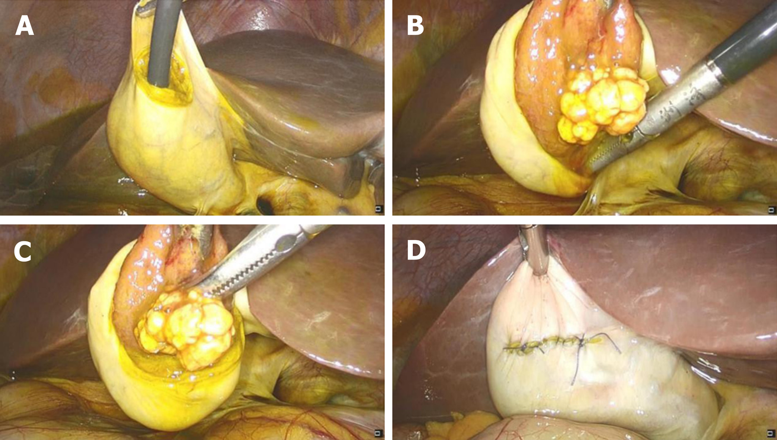
Figure 1 Laparoscopic images.
A: Laparoscopic cholecystectomy, placing choledochoscope in the gallbladder to observe the gallbladder polyp; B: Placing the polyp into the abdominal cavity; C: Electrocoagulation resection of the gallbladder polyp; D: Suturing the gallbladder layer by layer.
- Citation: Tang BF, Dang T, Wang QH, Chang ZH, Han WJ. Confocal laser endomicroscopy distinguishing benign and malignant gallbladder polyps during choledochoscopic gallbladder-preserving polypectomy: A case report. World J Clin Cases 2020; 8(24): 6358-6363
- URL: https://www.wjgnet.com/2307-8960/full/v8/i24/6358.htm
- DOI: https://dx.doi.org/10.12998/wjcc.v8.i24.6358


