Copyright
©The Author(s) 2020.
World J Clin Cases. Dec 6, 2020; 8(23): 6197-6205
Published online Dec 6, 2020. doi: 10.12998/wjcc.v8.i23.6197
Published online Dec 6, 2020. doi: 10.12998/wjcc.v8.i23.6197
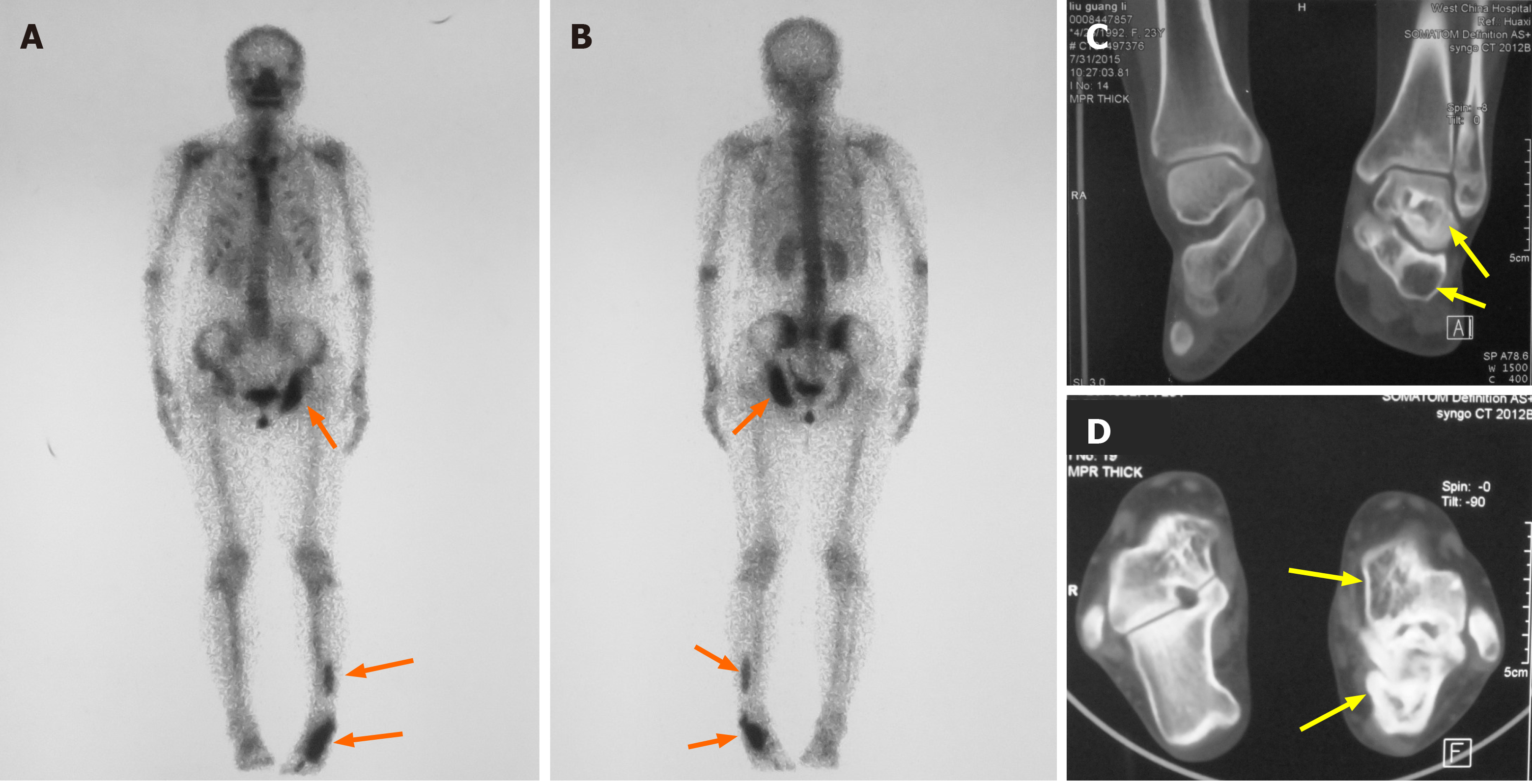
Figure 2 Radiography examination results.
A: Anterior view; B: Posterior view of skeletal scintigraphy showing the location of the bone lesions on left ischium and left fibula, talus, and calcaneus (orange arrows); C and D: Computed tomography shows well-circumscribed bone lucencies and ground-glass opacities in left talus and calcaneus (yellow arrows).
- Citation: Lin T, Li XY, Zou CY, Liu WW, Lin JF, Zhang XX, Zhao SQ, Xie XB, Huang G, Yin JQ, Shen JN. Discontinuous polyostotic fibrous dysplasia with multiple systemic disorders and unique genetic mutations: A case report. World J Clin Cases 2020; 8(23): 6197-6205
- URL: https://www.wjgnet.com/2307-8960/full/v8/i23/6197.htm
- DOI: https://dx.doi.org/10.12998/wjcc.v8.i23.6197


