
















©The Author(s) 2020.
World J Clin Cases. Dec 6, 2020; 8(23): 6190-6196
Published online Dec 6, 2020. doi: 10.12998/wjcc.v8.i23.6190
Published online Dec 6, 2020. doi: 10.12998/wjcc.v8.i23.6190
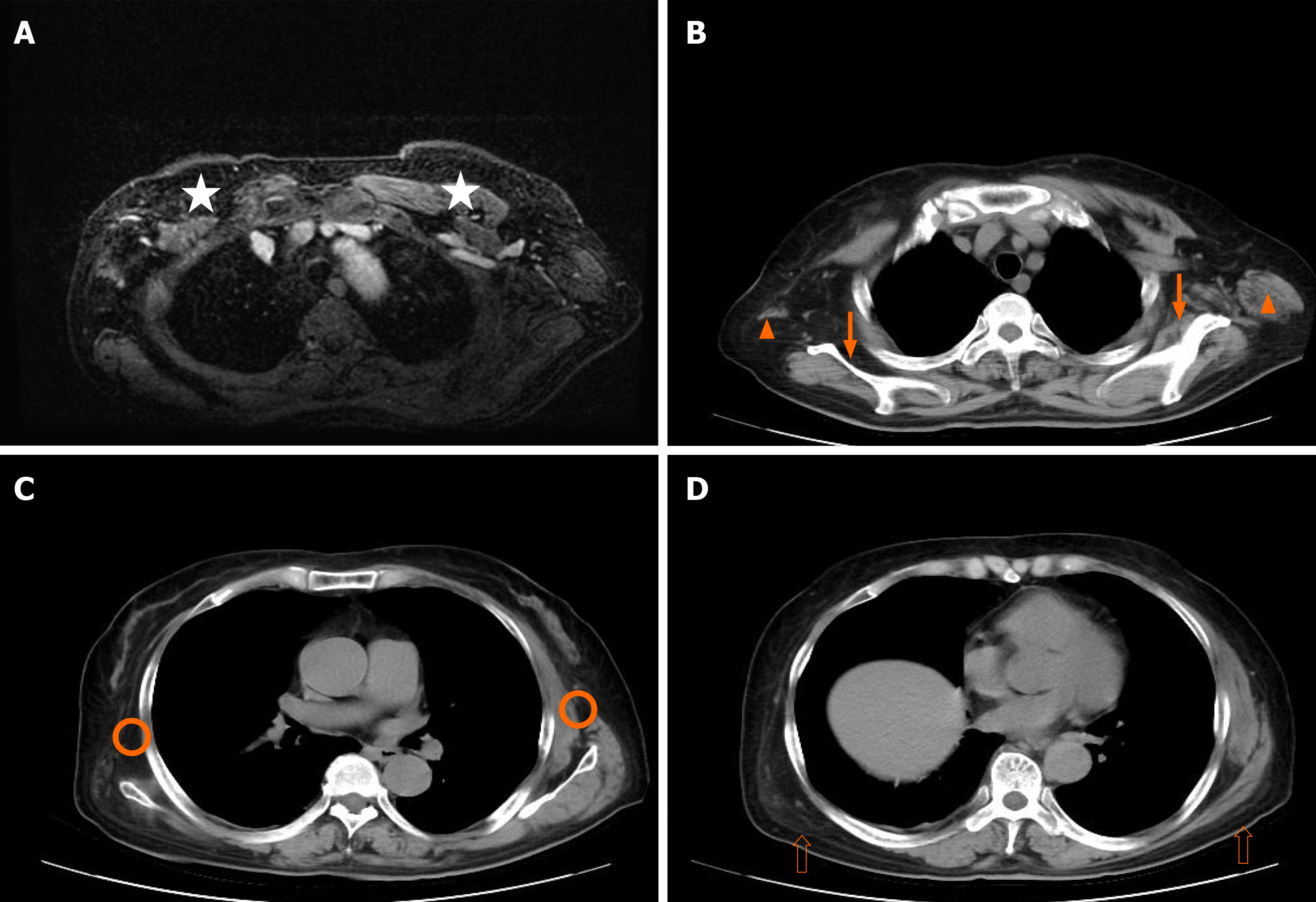
Figure 3 Chest imaging.
A: Axial magnetic resonance imaging; B, C, and D: CT scan of the chest revealed atrophy of the pectoralis major (star), teres major (orange triangle), serratus anterior (orange circle), subscapularis muscle (solid arrow), and latissimus dorsi muscle (hollow arrow).
- Citation: Wang XM, Cong YZ, Qiao GD, Zhang S, Wang LJ. Primary breast cancer patient with poliomyelitis: A case report. World J Clin Cases 2020; 8(23): 6190-6196
- URL: https://www.wjgnet.com/2307-8960/full/v8/i23/6190.htm
- DOI: https://dx.doi.org/10.12998/wjcc.v8.i23.6190