Copyright
©The Author(s) 2020.
World J Clin Cases. Nov 26, 2020; 8(22): 5751-5757
Published online Nov 26, 2020. doi: 10.12998/wjcc.v8.i22.5751
Published online Nov 26, 2020. doi: 10.12998/wjcc.v8.i22.5751
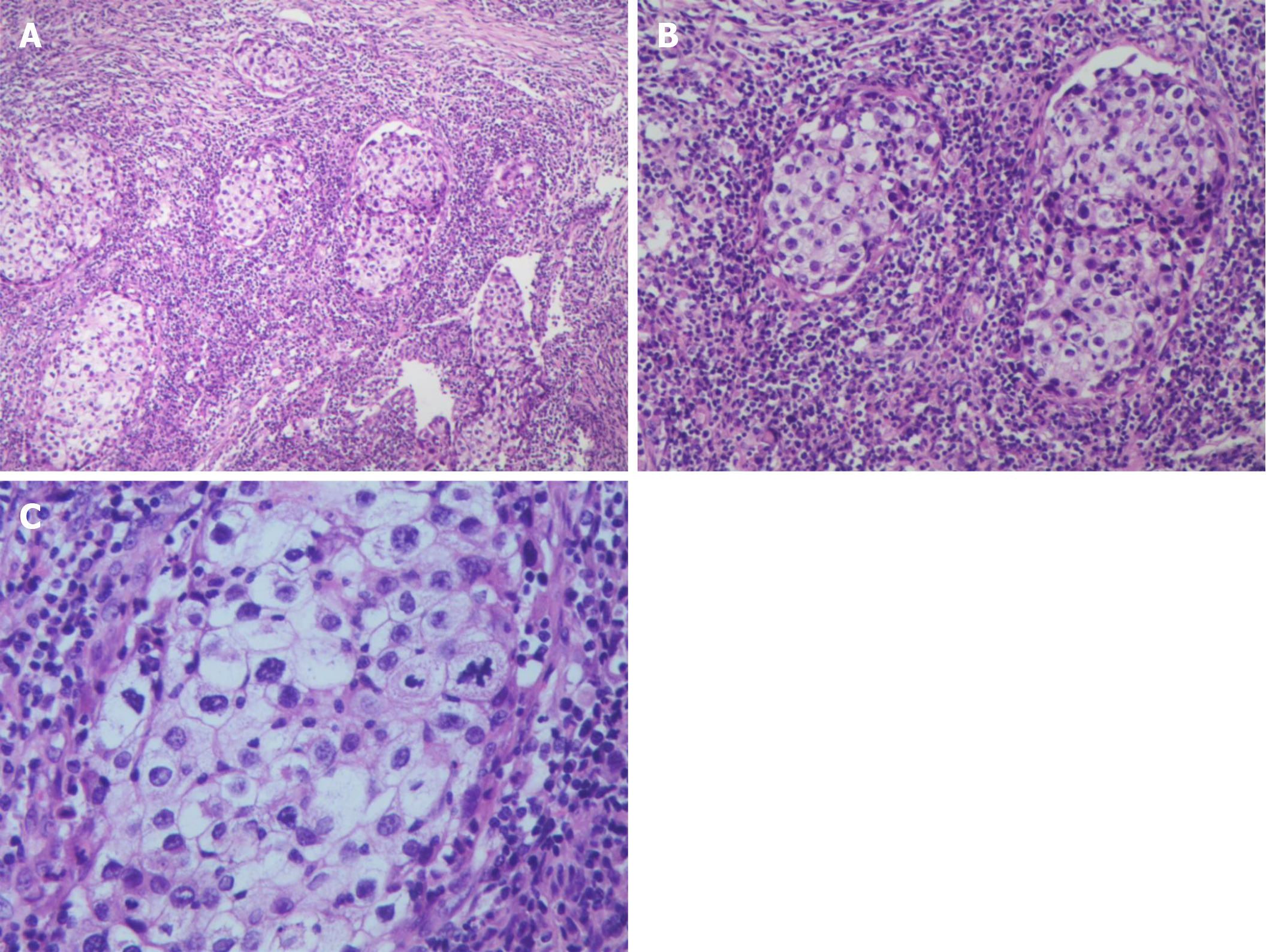
Figure 3 Hematoxylin and eosin staining and microscopic examination revealed a large number of lymphocytes and tumor cells with nest-like and lamellar infiltrative growth.
The tumor cells were rich in transparent to eosinophilic cytoplasm, with hyperchromatic nuclei and heterotypic and pathological nuclear division. A: × 100 magnification; B: × 200 magnification; C: × 400 magnification.
- Citation: Hao FY, Wang YL, Li SM, Xue LF. Sebaceous lymphadenocarcinoma of the parotid gland: A case report. World J Clin Cases 2020; 8(22): 5751-5757
- URL: https://www.wjgnet.com/2307-8960/full/v8/i22/5751.htm
- DOI: https://dx.doi.org/10.12998/wjcc.v8.i22.5751


