Copyright
©The Author(s) 2020.
World J Clin Cases. Nov 26, 2020; 8(22): 5701-5706
Published online Nov 26, 2020. doi: 10.12998/wjcc.v8.i22.5701
Published online Nov 26, 2020. doi: 10.12998/wjcc.v8.i22.5701
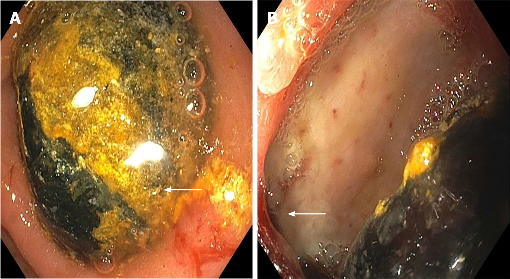
Figure 2 Esophagogastroduodenoscopy showing large impacted gall stone.
A: In the duodenal bulb; B: With an ulcerated cholecystoduodenal fistula (shown by arrows).
- Citation: Parvataneni S, Khara HS, Diehl DL. Bouveret syndrome masquerading as a gastric mass-unmasked with endoscopic luminal laser lithotripsy: A case report. World J Clin Cases 2020; 8(22): 5701-5706
- URL: https://www.wjgnet.com/2307-8960/full/v8/i22/5701.htm
- DOI: https://dx.doi.org/10.12998/wjcc.v8.i22.5701


