Copyright
©The Author(s) 2020.
World J Clin Cases. Nov 26, 2020; 8(22): 5625-5631
Published online Nov 26, 2020. doi: 10.12998/wjcc.v8.i22.5625
Published online Nov 26, 2020. doi: 10.12998/wjcc.v8.i22.5625
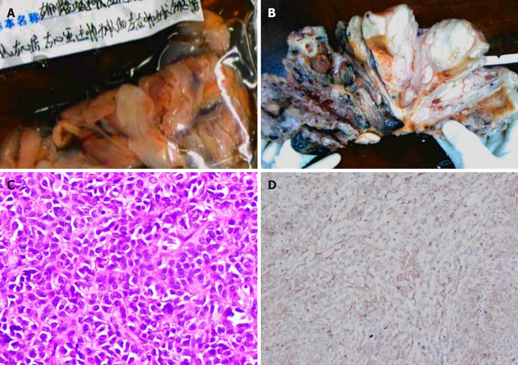
Figure 5 Pathological images.
A: Endovascular and intracardiac tumors; B: Cut section of the uterine tumor shows that the tumor diffusely infiltrated the whole layer of the uterine wall; C: Microscopically, the tumor cells were closely arranged and cytologically atypical (hematoxylin eosin staining, × 400); D: The tumor shows positive CD10 immunoreactivity.
- Citation: Fan JK, Tang GC, Yang H. Endometrial stromal sarcoma extending to the pulmonary artery: A rare case report. World J Clin Cases 2020; 8(22): 5625-5631
- URL: https://www.wjgnet.com/2307-8960/full/v8/i22/5625.htm
- DOI: https://dx.doi.org/10.12998/wjcc.v8.i22.5625


