Copyright
©The Author(s) 2020.
World J Clin Cases. Nov 26, 2020; 8(22): 5625-5631
Published online Nov 26, 2020. doi: 10.12998/wjcc.v8.i22.5625
Published online Nov 26, 2020. doi: 10.12998/wjcc.v8.i22.5625
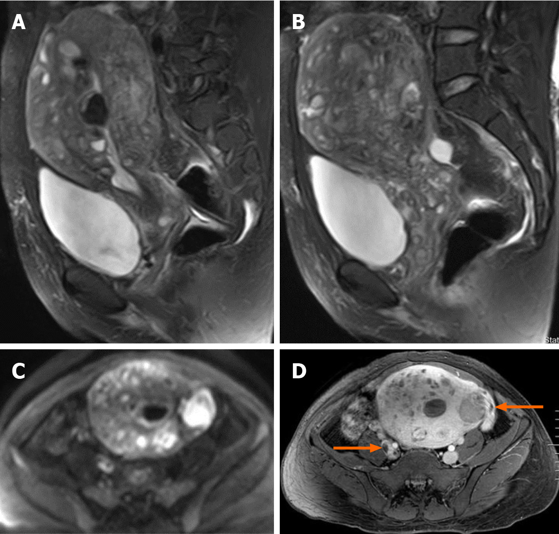
Figure 3 Pelvic magnetic resonance imaging findings.
A and B: Sagittal T2-weighted images shows extensive myometrial thickening with heterogeneously hyperintense signals, and diffuse infiltration of the cervix and vaginal vault; C: Axial diffusion-weighted image shows high lesion signals; D: Axial T1-weighted image after contrast administration shows that contrast enhancement of the lesions was heterogeneous and tumor thrombus in the right iliac vein and the left ovarian vein (arrow).
- Citation: Fan JK, Tang GC, Yang H. Endometrial stromal sarcoma extending to the pulmonary artery: A rare case report. World J Clin Cases 2020; 8(22): 5625-5631
- URL: https://www.wjgnet.com/2307-8960/full/v8/i22/5625.htm
- DOI: https://dx.doi.org/10.12998/wjcc.v8.i22.5625


