Copyright
©The Author(s) 2020.
World J Clin Cases. Nov 26, 2020; 8(22): 5625-5631
Published online Nov 26, 2020. doi: 10.12998/wjcc.v8.i22.5625
Published online Nov 26, 2020. doi: 10.12998/wjcc.v8.i22.5625
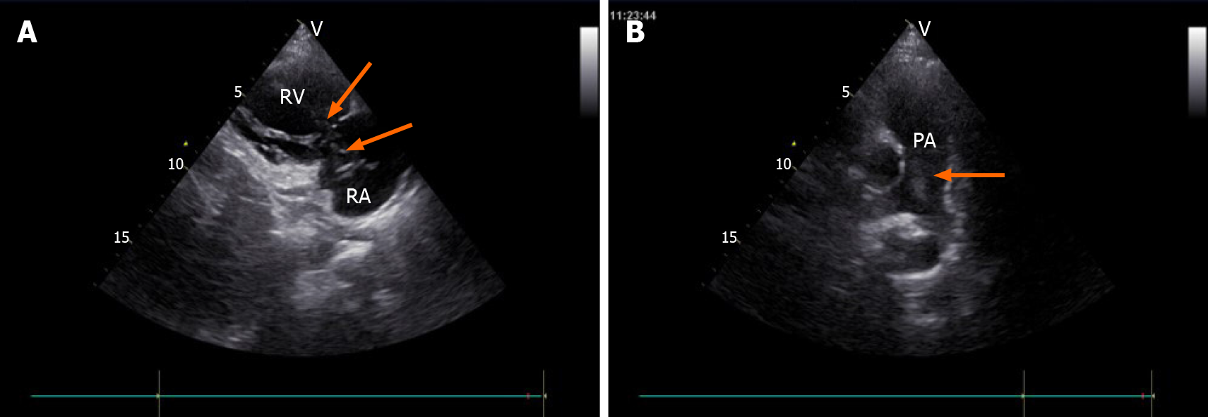
Figure 1 Transthoracic echocardiography images.
A: Tumor (arrow) in the right atrium and the right ventricle, seen on the parasternal right ventricular inflow tract section; B: Tumor (arrow) in the pulmonary artery, seen on the long axis section of the pulmonary artery. RA: Right atrium; RV: Right ventricle; PA: Pulmonary artery.
- Citation: Fan JK, Tang GC, Yang H. Endometrial stromal sarcoma extending to the pulmonary artery: A rare case report. World J Clin Cases 2020; 8(22): 5625-5631
- URL: https://www.wjgnet.com/2307-8960/full/v8/i22/5625.htm
- DOI: https://dx.doi.org/10.12998/wjcc.v8.i22.5625


