Copyright
©The Author(s) 2020.
World J Clin Cases. Nov 6, 2020; 8(21): 5326-5333
Published online Nov 6, 2020. doi: 10.12998/wjcc.v8.i21.5326
Published online Nov 6, 2020. doi: 10.12998/wjcc.v8.i21.5326
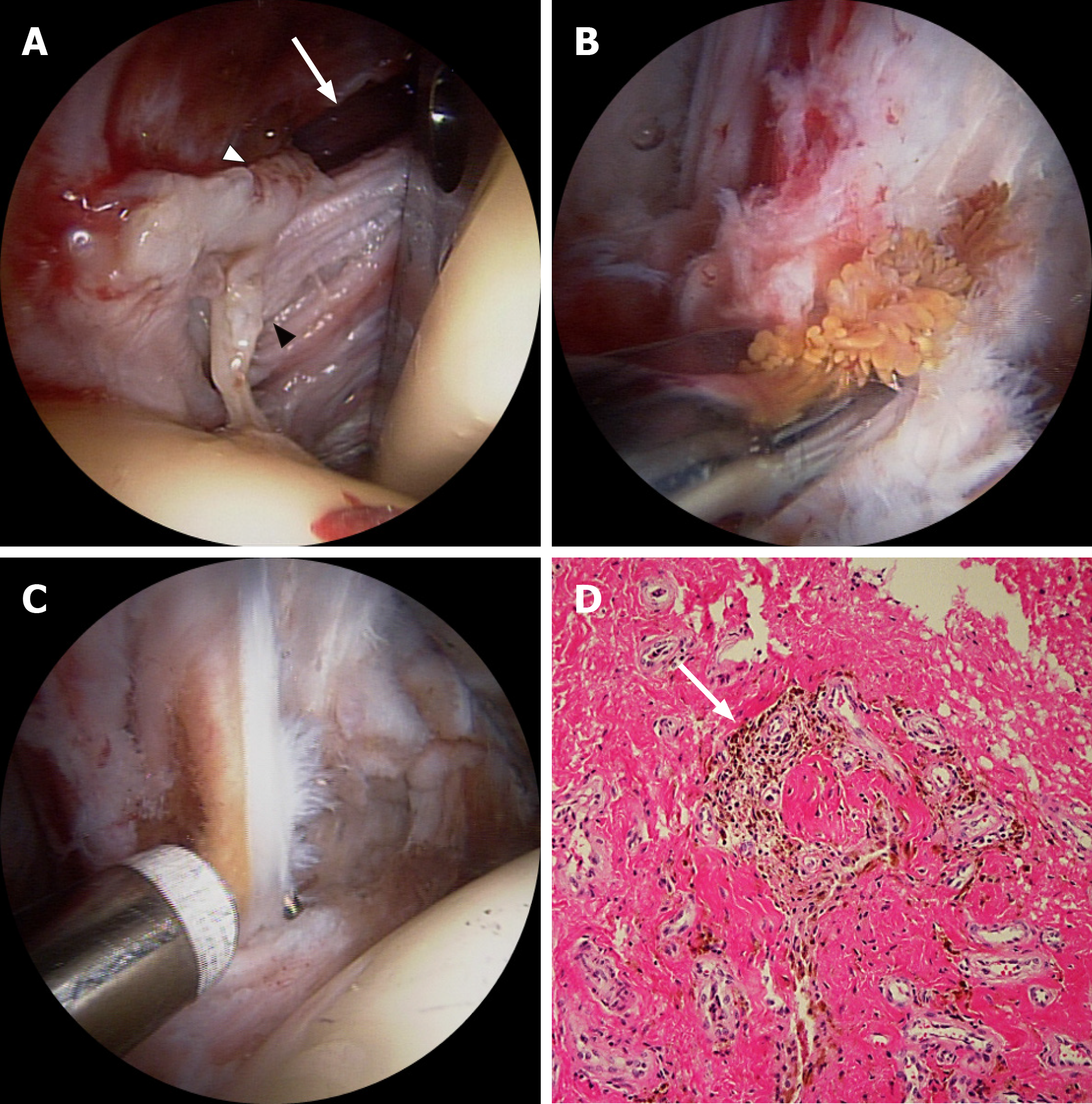
Figure 5 Arthroscopic findings and histological features in Case 1.
A: The capsular opening (white arrow) was enlarged and surrounded with frayed fibrous tissues (black arrowhead) and dark yellow amorphous material (white arrowhead), which looked like granulation tissue (anterolateral viewing portal); B: Partial synovectomy with biopsy (anterolateral viewing portal); C: Iliopsoas tendon release and bursectomy were performed (anterolateral viewing portal); D: Histopathologic study showed chronic inflammation with synovial hyperplasia and hemosiderin pigmentation (arrow; magnification, 200 ×).
- Citation: Won H, Kim KH, Jung JW, Kim SY, Baek SH. Arthroscopic treatment of iliopsoas tendinitis after total hip arthroplasty with acetabular cup malposition: Two case reports. World J Clin Cases 2020; 8(21): 5326-5333
- URL: https://www.wjgnet.com/2307-8960/full/v8/i21/5326.htm
- DOI: https://dx.doi.org/10.12998/wjcc.v8.i21.5326


