Copyright
©The Author(s) 2020.
World J Clin Cases. Nov 6, 2020; 8(21): 5326-5333
Published online Nov 6, 2020. doi: 10.12998/wjcc.v8.i21.5326
Published online Nov 6, 2020. doi: 10.12998/wjcc.v8.i21.5326
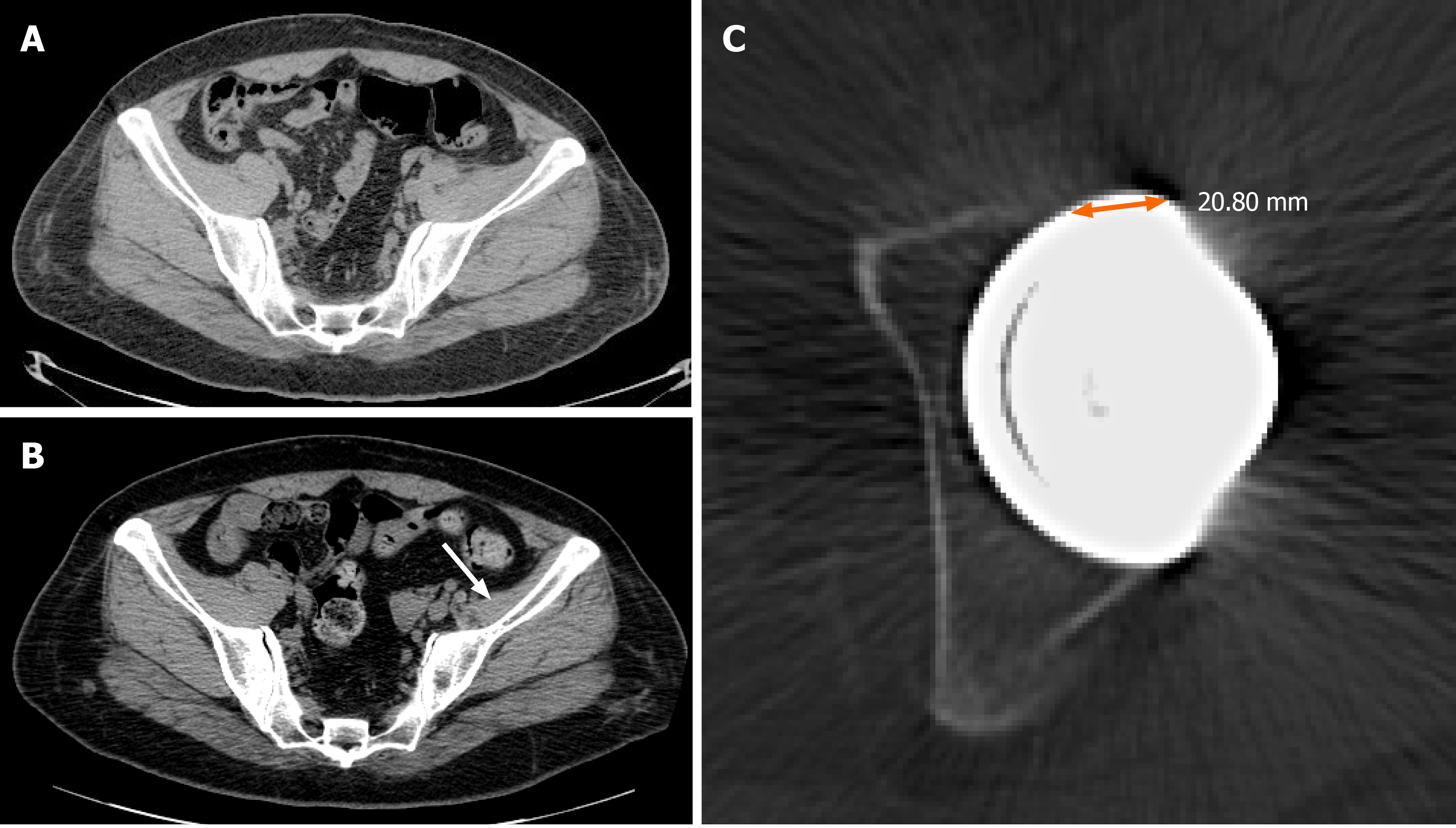
Figure 4 Computed tomography scans of Case 2.
A: Computed tomography (CT) scan taken immediately after perioperative dislocation to evaluate anteversion angle of acetabular cup and stem; B: CT scan taken 2 years after total hip arthroplasty, which demonstrated atrophy of the left iliacus muscle (arrow); C: Anteversion of the acetabular component, with an angle of 4 degrees and anterior prominence of more than 20 mm.
- Citation: Won H, Kim KH, Jung JW, Kim SY, Baek SH. Arthroscopic treatment of iliopsoas tendinitis after total hip arthroplasty with acetabular cup malposition: Two case reports. World J Clin Cases 2020; 8(21): 5326-5333
- URL: https://www.wjgnet.com/2307-8960/full/v8/i21/5326.htm
- DOI: https://dx.doi.org/10.12998/wjcc.v8.i21.5326


