Copyright
©The Author(s) 2020.
World J Clin Cases. Oct 26, 2020; 8(20): 5007-5012
Published online Oct 26, 2020. doi: 10.12998/wjcc.v8.i20.5007
Published online Oct 26, 2020. doi: 10.12998/wjcc.v8.i20.5007
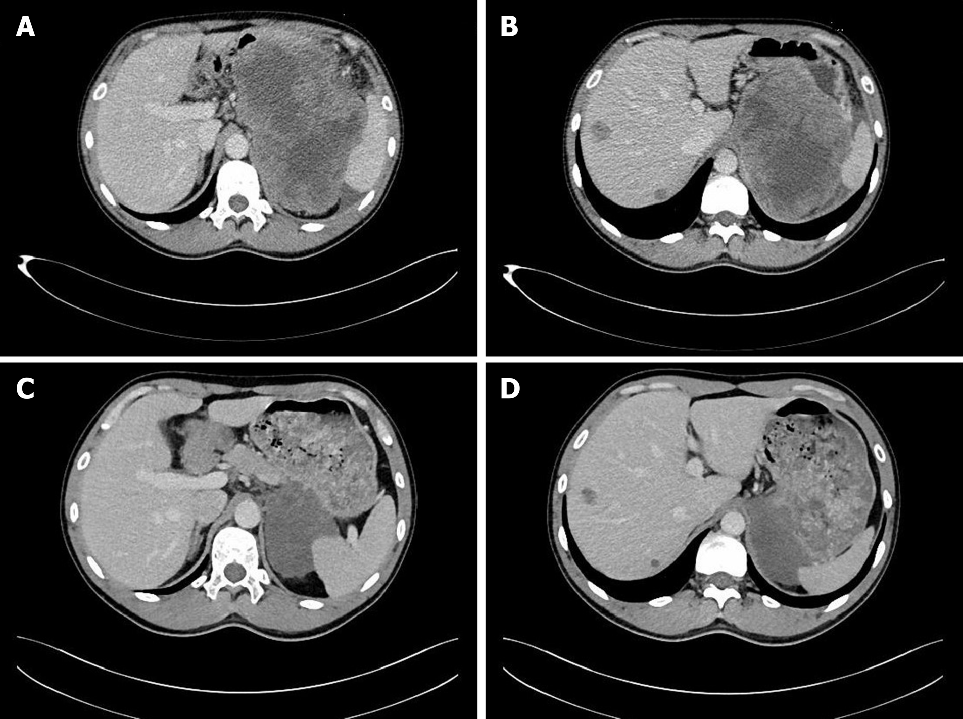
Figure 1 Computed tomography image.
A and B: Computed tomography (CT) showed a huge inhomogeneous soft tissue mass (approximately 18.1 cm × 11.9 cm) with central necrosis, occupying the entire left upper abdomen, along with two nodular enhancing liver lesions; C and D: CT after 10 mo of imatinib therapy showed marked reduction in tumor size (approximately 8.6 cm × 6.7 cm), tumor enhancement at arterial phase CT decreased substantially and hepatic metastatic lesions showed no enhancement.
- Citation: Yin XN, Yin Y, Wang J, Shen CY, Chen X, Zhao Z, Cai ZL, Zhang B. Gastrointestinal stromal tumor metastasis at the site of a totally implantable venous access port insertion: A rare case report. World J Clin Cases 2020; 8(20): 5007-5012
- URL: https://www.wjgnet.com/2307-8960/full/v8/i20/5007.htm
- DOI: https://dx.doi.org/10.12998/wjcc.v8.i20.5007


