Copyright
©The Author(s) 2020.
World J Clin Cases. Oct 26, 2020; 8(20): 4922-4929
Published online Oct 26, 2020. doi: 10.12998/wjcc.v8.i20.4922
Published online Oct 26, 2020. doi: 10.12998/wjcc.v8.i20.4922
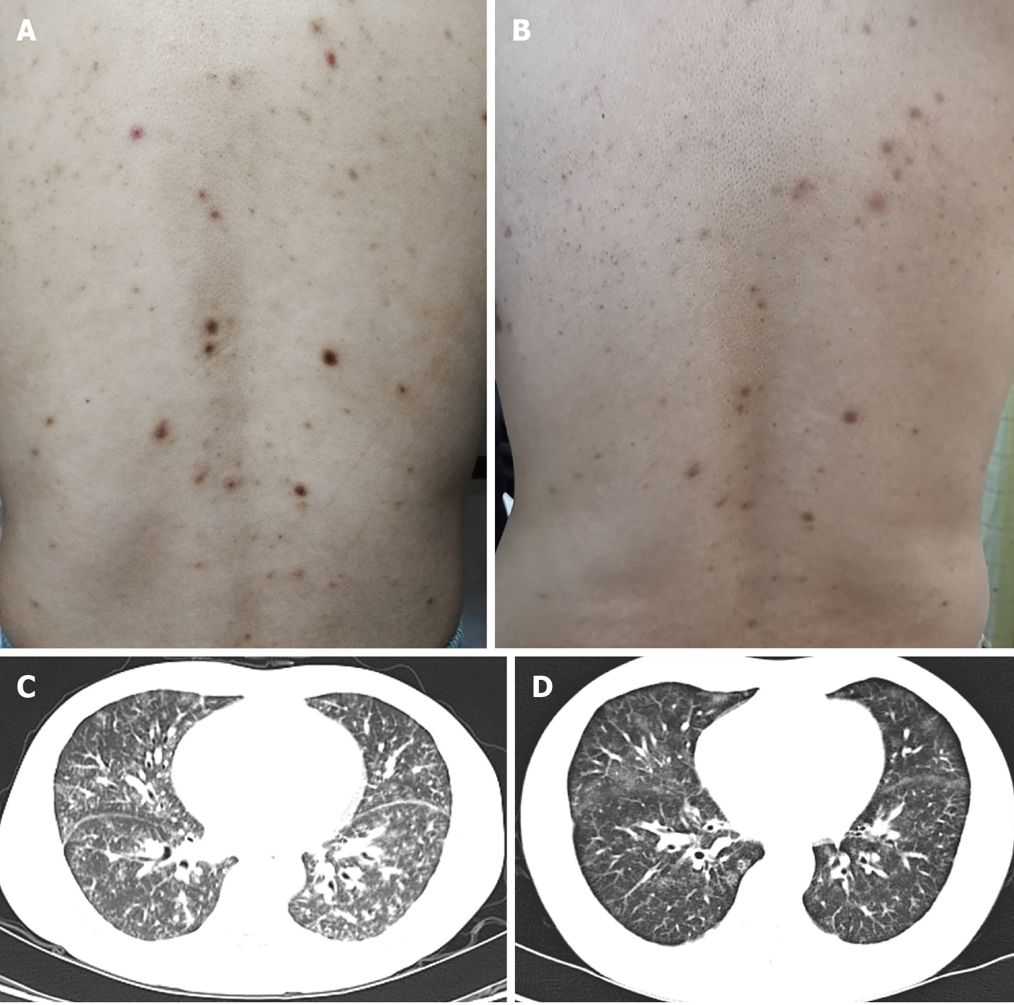
Figure 1 Skin lesions and chest computed tomography findings before and after treatment.
A: Innumerable symmetric brownish-red macules, papules, and plaques about 0.2-1 cm in diameter distributed in the back before treatment; B: The skin lesion improved 9 mo after treatment; C: Parenchymal window setting of the chest computed tomography showed diffuse ground-glass opacities in two lobes before treatment; D: The lung lesion improved 9 mo after treatment.
- Citation: Han PY, Chi HH, Su YT. Idiopathic multicentric Castleman disease with pulmonary and cutaneous lesions treated with tocilizumab: A case report . World J Clin Cases 2020; 8(20): 4922-4929
- URL: https://www.wjgnet.com/2307-8960/full/v8/i20/4922.htm
- DOI: https://dx.doi.org/10.12998/wjcc.v8.i20.4922


