Copyright
©The Author(s) 2020.
World J Clin Cases. Oct 26, 2020; 8(20): 4753-4762
Published online Oct 26, 2020. doi: 10.12998/wjcc.v8.i20.4753
Published online Oct 26, 2020. doi: 10.12998/wjcc.v8.i20.4753
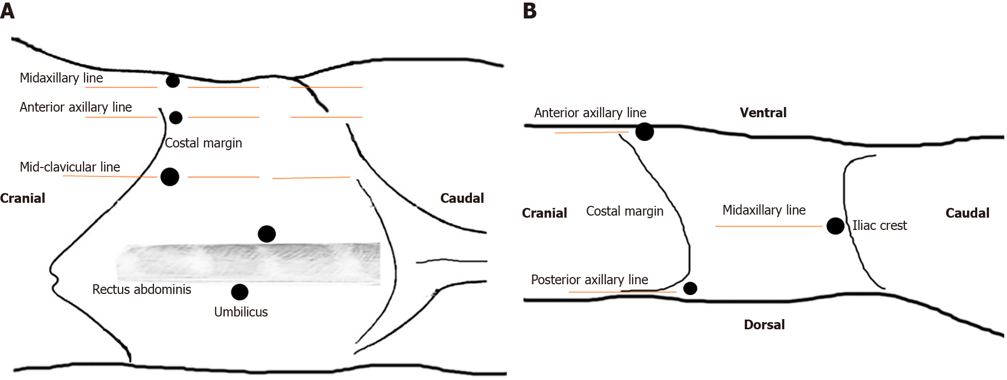
Figure 1 The trocar position during laparoscopic treatment.
A: Transperitoneal procedure: Two 10-mm trocars were placed, one at the umbilicus and the other 2 cm below the umbilicus adjacent to the rectus sheath, respectively. The trocar at the umbilicus was the camera port. A 12-mm trocar was inserted 2 cm below the costal margin in the mid-clavicular line. The other two 5-mm trocars were placed 2 cm below the costal margin in the anterior axillary line and midaxillary line, respectively; B: Retroperitoneal procedure: One 10-mm trocar was located 1 cm above the level of the iliac crest in the midaxillary line as the camera port. One 12-mm trocar was placed 2 cm below the costal margin in the anterior axillary line. One 5-mm trocar was inserted 1 cm below the costal margin in the posterior axillary line.
- Citation: Chen X, Wang Y, Gao L, Song J, Wang JY, Wang DD, Ma JX, Zhang ZQ, Bi LK, Xie DD, Yu DX. Retroperitoneal vs transperitoneal laparoscopic lithotripsy of 20-40 mm renal stones within horseshoe kidneys. World J Clin Cases 2020; 8(20): 4753-4762
- URL: https://www.wjgnet.com/2307-8960/full/v8/i20/4753.htm
- DOI: https://dx.doi.org/10.12998/wjcc.v8.i20.4753


