
















©The Author(s) 2020.
World J Clin Cases. Jan 26, 2020; 8(2): 471-478
Published online Jan 26, 2020. doi: 10.12998/wjcc.v8.i2.471
Published online Jan 26, 2020. doi: 10.12998/wjcc.v8.i2.471
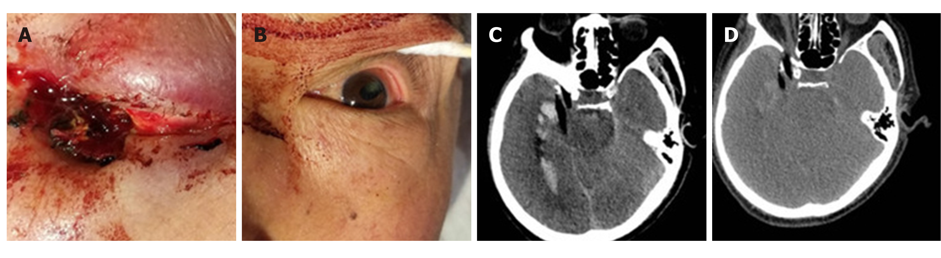
Figure 1 Physical and imaging examinations of case 1.
A: An exposed wooden foreign body, 0.8 cm in diameter, was observed in the right inferior orbital wall accompanied by blood exudation; B: The left pupil, with a history of trauma, was elliptic, with a long diameter of 5.0 mm and a short diameter of 3.0 mm; C and D: Head computed tomography showed a strip of low-density shadow under the right eyeball, entering the brain through the orbital apex and lateral orbital apex. The optic nerve was compressed. The lateral rectus was distorted and displaced.
- Citation: Xue H, Zhang WT, Wang GM, Shi L, Zhang YM, Yang HF. Transorbital nonmissile penetrating brain injury: Report of two cases. World J Clin Cases 2020; 8(2): 471-478
- URL: https://www.wjgnet.com/2307-8960/full/v8/i2/471.htm
- DOI: https://dx.doi.org/10.12998/wjcc.v8.i2.471