
















©The Author(s) 2020.
World J Clin Cases. Jan 26, 2020; 8(2): 370-376
Published online Jan 26, 2020. doi: 10.12998/wjcc.v8.i2.370
Published online Jan 26, 2020. doi: 10.12998/wjcc.v8.i2.370
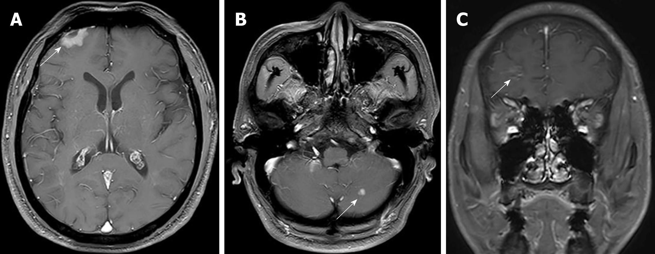
Figure 1 Baseline axial (A and B) and sagittal (C) T1w C+ craniocerebral magnetic resonance imaging showed the central nervous system metastases in the right frontal lobe (brain metastases and leptomeningeal metastases) and left cerebellar hemisphere (brain metastases) (white arrows).
- Citation: Li HY, Xie Y, Yu TT, Lin YJ, Yin ZY. Durable response to pulsatile icotinib for central nervous system metastases from EGFR-mutated non-small cell lung cancer: A case report. World J Clin Cases 2020; 8(2): 370-376
- URL: https://www.wjgnet.com/2307-8960/full/v8/i2/370.htm
- DOI: https://dx.doi.org/10.12998/wjcc.v8.i2.370