Copyright
©The Author(s) 2020.
World J Clin Cases. Jan 26, 2020; 8(2): 325-330
Published online Jan 26, 2020. doi: 10.12998/wjcc.v8.i2.325
Published online Jan 26, 2020. doi: 10.12998/wjcc.v8.i2.325
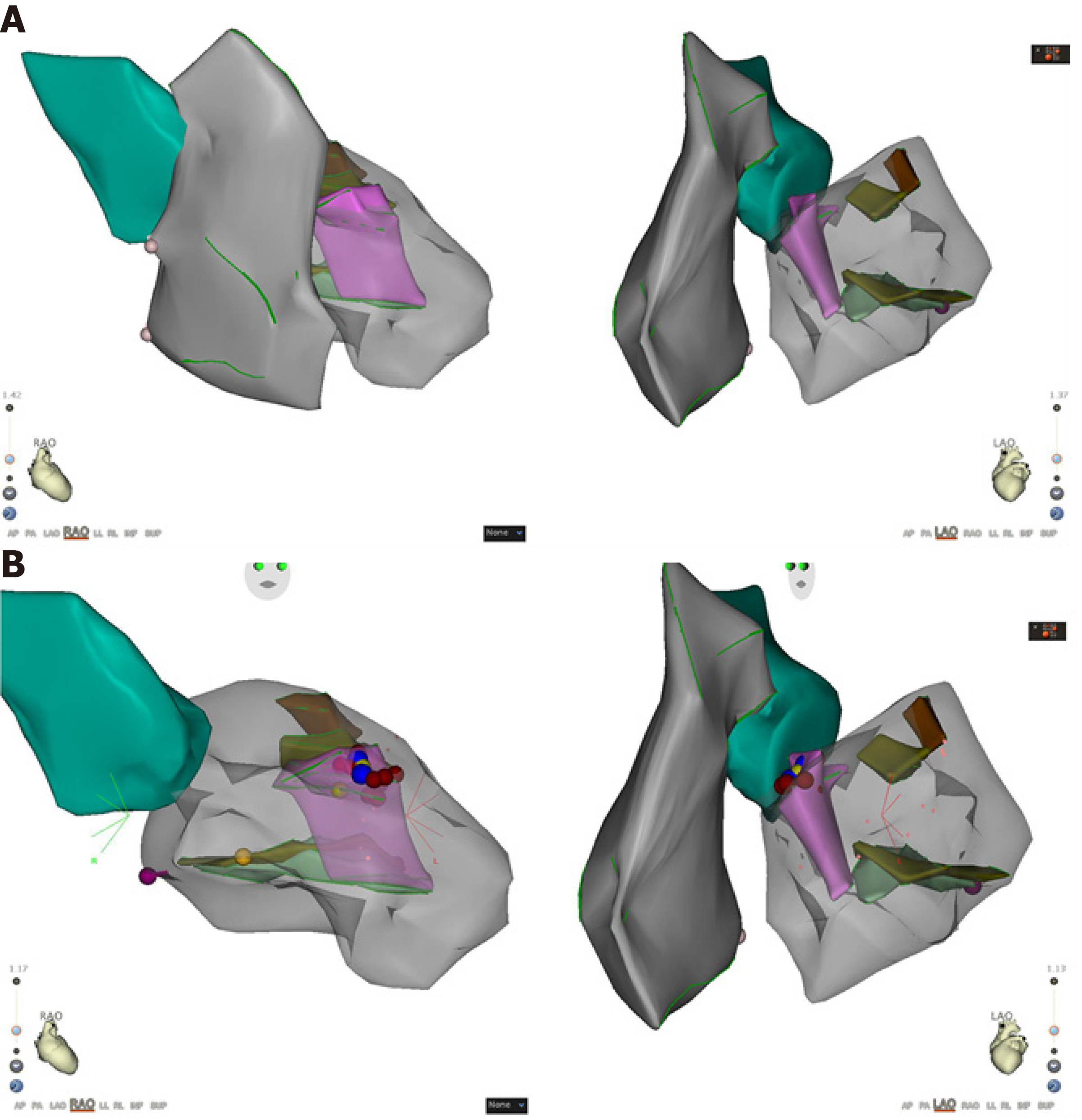
Figure 3 Three-dimensional structure model.
A: Left ventricular model established with an intracardiac ultrasound catheter and target map marked by the ST catheter in the left ventricle. In the model, pink represents a false tendon, brown represents the anterior papillary muscle, and green represents the posterior papillary muscle; B: The three-dimensional model during ablation and intracardiac ultrasound indicates that the target point is located at the attachment of the false tendon near the basal side of the interventricular septum.
- Citation: Yang YB, Li XF, Guo TT, Jia YH, Liu J, Tang M, Fang PH, Zhang S. Catheter ablation of premature ventricular complexes associated with false tendons: A case report. World J Clin Cases 2020; 8(2): 325-330
- URL: https://www.wjgnet.com/2307-8960/full/v8/i2/325.htm
- DOI: https://dx.doi.org/10.12998/wjcc.v8.i2.325


